Copyright
©2005 Baishideng Publishing Group Inc.
World J Gastroenterol. Apr 28, 2005; 11(16): 2402-2407
Published online Apr 28, 2005. doi: 10.3748/wjg.v11.i16.2402
Published online Apr 28, 2005. doi: 10.3748/wjg.v11.i16.2402
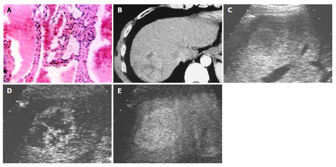
Figure 3 A: a 4.
1 cm-sized hemangioma; B: heterogenous high attenuation on contrast-enhanced CT; C: hypoechoic lesion on B-mode image; D: the spot-like enhancement in early arterial phase on contrast-enhanced C3-MODE technology; E: centripetal filling in late phase (e) on contrast-enhanced C3-MODE technology.
- Citation: Luo BM, Wen YL, Yang HY, Zhi H, Ou B, Ma JH, Pan JS, Dai XN. Differentiation between malignant and benign nodules in the liver: Use of contrast C3-MODE technology. World J Gastroenterol 2005; 11(16): 2402-2407
- URL: https://www.wjgnet.com/1007-9327/full/v11/i16/2402.htm
- DOI: https://dx.doi.org/10.3748/wjg.v11.i16.2402


