Copyright
©2005 Baishideng Publishing Group Inc.
World J Gastroenterol. Apr 14, 2005; 11(14): 2188-2192
Published online Apr 14, 2005. doi: 10.3748/wjg.v11.i14.2188
Published online Apr 14, 2005. doi: 10.3748/wjg.v11.i14.2188
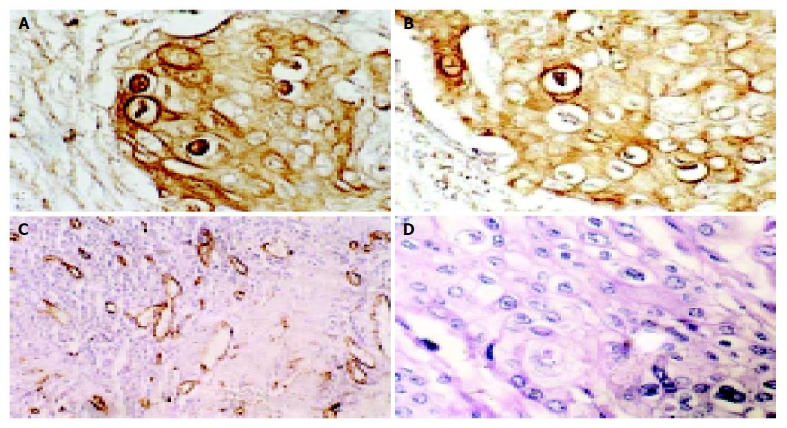
Figure 1 Immunohistochemistry SP staining and HE staining A: Heparanase-positive (nucleolus, cytoplasm, amphithecium) SP×400 B: bFGF-heparanase-positive (cytoplasm, amphithecium) SP×400 C: MVD as evaluated by immunohistochemistry with CD34 (SPx200) D: HE staining of EC SP×400.
- Citation: Han B, Liu J, Ma MJ, Zhao L. Clinicopathological significance of heparanase and basic fibroblast growth factor expression in human esophageal cancer. World J Gastroenterol 2005; 11(14): 2188-2192
- URL: https://www.wjgnet.com/1007-9327/full/v11/i14/2188.htm
- DOI: https://dx.doi.org/10.3748/wjg.v11.i14.2188


