Copyright
©2005 Baishideng Publishing Group Inc.
World J Gastroenterol. Apr 14, 2005; 11(14): 2101-2108
Published online Apr 14, 2005. doi: 10.3748/wjg.v11.i14.2101
Published online Apr 14, 2005. doi: 10.3748/wjg.v11.i14.2101
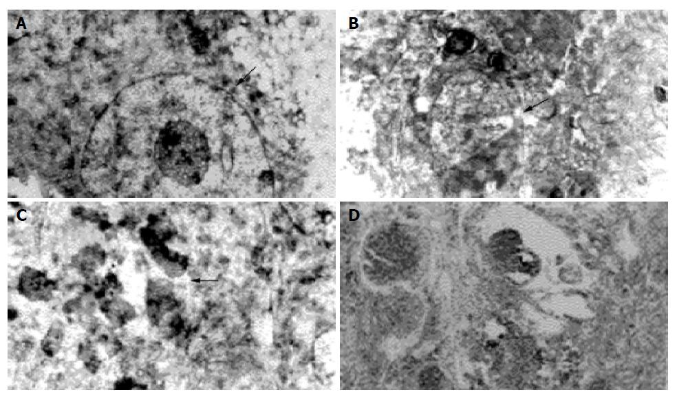
Figure 4 Transmission electron microscopy of ILN A: Metastatic Pc-3 cells (arrow) in lymphatic tissue of control group (4×10³); B: After 32P colloids irradiation apoptotic cells (arrow) in the connective tissue and endothelium of lymphatic sinuses (2.
5×10³); C: Denatured tumor cells (arrow) after medication (5×103×1.2); D: ILN under light microscope: lymphatic tissue recovered to normal 4 wk after medication (HE ×400).
- Citation: Liu L, Feng GS, Gao H, Tong GS, Wang Y, Gao W, Huang Y, Li C. Chromic-P32 phosphate treatment of implanted pancreatic carcinoma: Mechanism involved. World J Gastroenterol 2005; 11(14): 2101-2108
- URL: https://www.wjgnet.com/1007-9327/full/v11/i14/2101.htm
- DOI: https://dx.doi.org/10.3748/wjg.v11.i14.2101


