Copyright
©2005 Baishideng Publishing Group Inc.
World J Gastroenterol. Apr 7, 2005; 11(13): 1896-1902
Published online Apr 7, 2005. doi: 10.3748/wjg.v11.i13.1896
Published online Apr 7, 2005. doi: 10.3748/wjg.v11.i13.1896
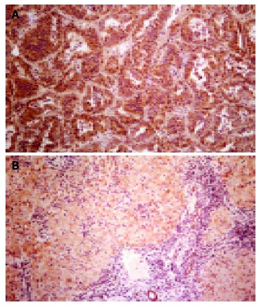
Figure 1 Immunohistochemical staining for COX-2.
A: a section of a HCC showing expression of the COX-2 protein (brownish staining) predominantly in the tumor cells; B: a section of nontumorous liver tissue showing expression of the COX-2 protein in the hepatocytes (magnification 10×).
- Citation: Tang TC, Poon RT, Lau CP, Xie D, Fan ST. Tumor cyclooxygenase-2 levels correlate with tumor invasiveness in human hepatocellular carcinoma. World J Gastroenterol 2005; 11(13): 1896-1902
- URL: https://www.wjgnet.com/1007-9327/full/v11/i13/1896.htm
- DOI: https://dx.doi.org/10.3748/wjg.v11.i13.1896


