Copyright
©2005 Baishideng Publishing Group Inc.
World J Gastroenterol. Mar 28, 2005; 11(12): 1881-1883
Published online Mar 28, 2005. doi: 10.3748/wjg.v11.i12.1881
Published online Mar 28, 2005. doi: 10.3748/wjg.v11.i12.1881
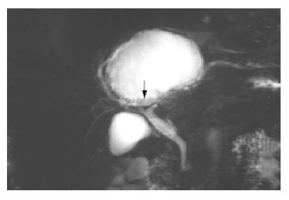
Figure 3 A T2-weighted MR image reveals a mass consisting mostly of fluid density.
The lower portion has low signal intensity (arrow), suggesting cyst hemorrhage or a cystic tumor.
- Citation: Lin CC, Lin SC, Ko WC, Chang KM, Shih SC. Adenocarcinoma and infection in a solitary hepatic cyst: A case report. World J Gastroenterol 2005; 11(12): 1881-1883
- URL: https://www.wjgnet.com/1007-9327/full/v11/i12/1881.htm
- DOI: https://dx.doi.org/10.3748/wjg.v11.i12.1881


