Copyright
©2005 Baishideng Publishing Group Inc.
World J Gastroenterol. Mar 28, 2005; 11(12): 1825-1828
Published online Mar 28, 2005. doi: 10.3748/wjg.v11.i12.1825
Published online Mar 28, 2005. doi: 10.3748/wjg.v11.i12.1825
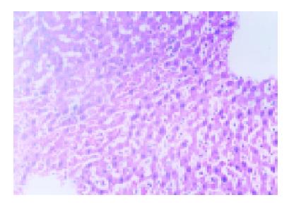
Figure 2 Liver tissue from I/R group showed disorderly liver sinusoids enlarged and congested with many red blood cells, lining endothelial cells necrotized and sloughed off, and multiple hepatocellular necrosis and massive infiltration of neutriphils as well.
HE×200.
- Citation: Yuan GJ, Ma JC, Gong ZJ, Sun XM, Zheng SH, Li X. Modulation of liver oxidant-antioxidant system by ischemic preconditioning during ischemia/reperfusion injury in rats. World J Gastroenterol 2005; 11(12): 1825-1828
- URL: https://www.wjgnet.com/1007-9327/full/v11/i12/1825.htm
- DOI: https://dx.doi.org/10.3748/wjg.v11.i12.1825


