Copyright
©2005 Baishideng Publishing Group Inc.
World J Gastroenterol. Mar 28, 2005; 11(12): 1788-1792
Published online Mar 28, 2005. doi: 10.3748/wjg.v11.i12.1788
Published online Mar 28, 2005. doi: 10.3748/wjg.v11.i12.1788
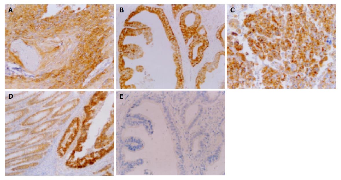
Figure 1 Expression and location of p62 detected by IHC.
EnVision (A–C, E×200, D×100). A: p62 expression in cytoplasm of squamous cell carcinoma but not in the site of keratinization; B: p62 expression in cytoplasm of well-differentiated adenocarcinoma; C: p62 expression in cytoplasm of HCC; D: Distribution of p62 in cytoplasm of morphologically normal columnar epithelial cells adjacent to cancer foci; E: Negative control.
- Citation: Qian HL, Peng XX, Chen SH, Ye HM, Qiu JH. p62 expression in primary carcinomas of the digestive system. World J Gastroenterol 2005; 11(12): 1788-1792
- URL: https://www.wjgnet.com/1007-9327/full/v11/i12/1788.htm
- DOI: https://dx.doi.org/10.3748/wjg.v11.i12.1788


