Copyright
©2005 Baishideng Publishing Group Inc.
World J Gastroenterol. Mar 21, 2005; 11(11): 1728-1729
Published online Mar 21, 2005. doi: 10.3748/wjg.v11.i11.1728
Published online Mar 21, 2005. doi: 10.3748/wjg.v11.i11.1728
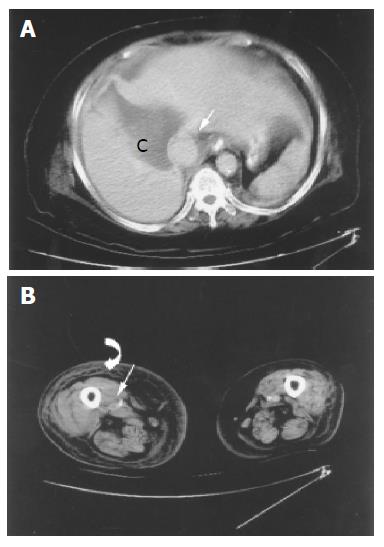
Figure 3 A: The post-drainage follow-up CT scan shows remarkable shrinkage of the hepatic cyst (C), no thrombi of IVC at this level (straight arrow).
B: However, there is still long segmental thrombi occupation of lower trunk of IVC and right lower leg, including right popliteal vein (straight arrow) and subcutaneous tissue swelling of lower extremity due to cellulitis (curve arrow).
- Citation: Leung TK, Lee CM, Chen HC. Fatal thrombotic complications of hepatic cystic compression of the inferior vena: A case report. World J Gastroenterol 2005; 11(11): 1728-1729
- URL: https://www.wjgnet.com/1007-9327/full/v11/i11/1728.htm
- DOI: https://dx.doi.org/10.3748/wjg.v11.i11.1728


