Copyright
©2005 Baishideng Publishing Group Inc.
World J Gastroenterol. Mar 21, 2005; 11(11): 1610-1615
Published online Mar 21, 2005. doi: 10.3748/wjg.v11.i11.1610
Published online Mar 21, 2005. doi: 10.3748/wjg.v11.i11.1610
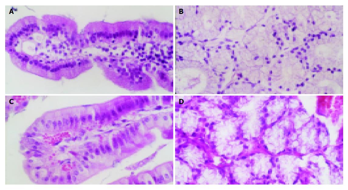
Figure 6 HE stain of duodenal structure of rabbits.
A: HE stain of normal rabbit’s duodenal villus. The minute blood vessel had not dilated and red blood cells had not leaked out from capillary lumens; B: HE stain of normal rabbit’s mucous glands of duodenum. The minute blood vessel had not dilated; C: HE stain of the rabbit’s duodenal villus, which were 48 h after brainstem hemorrhage. The capillaries had obviously dilated and red blood cells had leaked out from capillary lumens; D: HE stain of the rabbit’s mucous glands of duodenum, which were 48 h after brainstem hemorrhage. The capillaries had obviously dilated and red blood cells had leaked out from capillary lumens.
- Citation: Jin XL, Zheng Y, Shen HM, Jing WL, Zhang ZQ, Huang JZ, Tan QL. Analysis of the mechanisms of rabbit’s brainstem hemorrhage complicated with irritable changes in the alvine mucous membrane. World J Gastroenterol 2005; 11(11): 1610-1615
- URL: https://www.wjgnet.com/1007-9327/full/v11/i11/1610.htm
- DOI: https://dx.doi.org/10.3748/wjg.v11.i11.1610


