Copyright
©2005 Baishideng Publishing Group Inc.
World J Gastroenterol. Mar 21, 2005; 11(11): 1610-1615
Published online Mar 21, 2005. doi: 10.3748/wjg.v11.i11.1610
Published online Mar 21, 2005. doi: 10.3748/wjg.v11.i11.1610
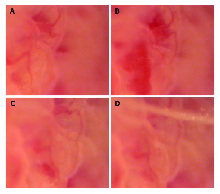
Figure 3 Changes of microcirculation of jejunal mucosa.
A: Photograph of microcirculation of jejunal mucous membrane, 6 s before the load of rabbit’s brainstem hemorrhage (×185); B: Photograph of jejunal mucous membrane, 10 min 26 s after the load of rabbit’s brainstem hemorrhage; it showed the local congestion of villus mucosa (×185); C: Photograph of jejunal mucous membrane, 30 min 32 s after the load of rabbit’s brainstem hemorrhage; it showed that the local congestion of villus mucosa had relieved (×185); D: Photograph of jejunal mucous membrane, 40 min 46 s after the load of rabbit’s brainstem hemorrhage; it showed that the local congestion of villus mucosa had ameliorated obviously (×185).
- Citation: Jin XL, Zheng Y, Shen HM, Jing WL, Zhang ZQ, Huang JZ, Tan QL. Analysis of the mechanisms of rabbit’s brainstem hemorrhage complicated with irritable changes in the alvine mucous membrane. World J Gastroenterol 2005; 11(11): 1610-1615
- URL: https://www.wjgnet.com/1007-9327/full/v11/i11/1610.htm
- DOI: https://dx.doi.org/10.3748/wjg.v11.i11.1610


