Copyright
©2005 Baishideng Publishing Group Inc.
World J Gastroenterol. Mar 14, 2005; 11(10): 1426-1432
Published online Mar 14, 2005. doi: 10.3748/wjg.v11.i10.1426
Published online Mar 14, 2005. doi: 10.3748/wjg.v11.i10.1426
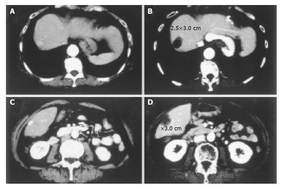
Figure 5 Two cases with small-sized HCCs treated with PEI-RFA under low power output control are shown.
Contrast-enhanced CT before (A, C) and after (B, D) PEI-RFA. Small HCCs of 1.5 cm in the longest diameter were located in the S8 region of the liver in both cases. In both cases, RFA was performed at 40 W for 5 min. Though the ablation was performed at relatively low power output for short duration, the coagulated necrosis larger than 2.5 cm was induced after the treatment.
- Citation: Kurokohchi K, Watanabe S, Masaki T, Hosomi N, Miyauchi Y, Himoto T, Kimura Y, Nakai S, Deguchi A, Yoneyama H, Yoshida S, Kuriyama S. Comparison between combination therapy of percutaneous ethanol injection and radiofrequency ablation and radiofrequency ablation alone for patients with hepatocellular carcinoma. World J Gastroenterol 2005; 11(10): 1426-1432
- URL: https://www.wjgnet.com/1007-9327/full/v11/i10/1426.htm
- DOI: https://dx.doi.org/10.3748/wjg.v11.i10.1426


