Copyright
©2005 Baishideng Publishing Group Inc.
World J Gastroenterol. Mar 14, 2005; 11(10): 1426-1432
Published online Mar 14, 2005. doi: 10.3748/wjg.v11.i10.1426
Published online Mar 14, 2005. doi: 10.3748/wjg.v11.i10.1426
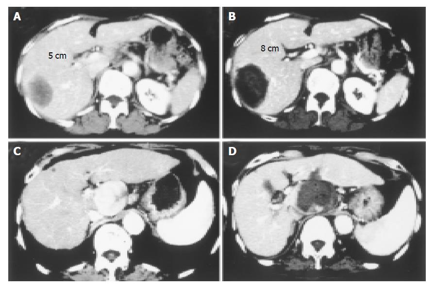
Figure 4 Two cases with large-sized HCC treated with PEI-RFA are shown.
Contrast-enhanced CT before (A: delay phase, C: early vascular phase) and after (B: delay phase, D: delay phase) PEI-RFA. Massive HCCs of 5 cm in the longest diameter were located in the right lobe of the liver in both cases. In the first case (A and B), RFA was started at 30 W and the power output was increased stepwise to 100 W every two min and the ablation was performed for 20 min. In the second case (C and D), because the tumor was located close by blood vessel such as inferior vena cava, portal tract and aorta, it was likely to be difficult to treat with RFA under high power control. After injecting 19 mL of ethanol into the tumor, one session of RFA was performed at 30 W for 12 min. The massive tumor was completely eliminated by PEI-RFA.
- Citation: Kurokohchi K, Watanabe S, Masaki T, Hosomi N, Miyauchi Y, Himoto T, Kimura Y, Nakai S, Deguchi A, Yoneyama H, Yoshida S, Kuriyama S. Comparison between combination therapy of percutaneous ethanol injection and radiofrequency ablation and radiofrequency ablation alone for patients with hepatocellular carcinoma. World J Gastroenterol 2005; 11(10): 1426-1432
- URL: https://www.wjgnet.com/1007-9327/full/v11/i10/1426.htm
- DOI: https://dx.doi.org/10.3748/wjg.v11.i10.1426


