Copyright
©The Author(s) 2004.
World J Gastroenterol. Apr 15, 2004; 10(8): 1208-1211
Published online Apr 15, 2004. doi: 10.3748/wjg.v10.i8.1208
Published online Apr 15, 2004. doi: 10.3748/wjg.v10.i8.1208
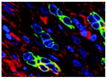
Figure 8 HLC.
Immunofluorencence confocal laser scanning microscopy (ICLSM) shows that two proliferating bile ductules are composed only of bile epithelial cells (anit CK7, green), but one proliferating bile ductule contains HPC (anti CK7 and albumin, orange). × 1 000.
- Citation: Xiao JC, Jin XL, Ruck P, Adam A, Kaiserling E. Hepatic progenitor cells in human liver cirrhosis: Immunohistochemical, electron microscopic and immunofluorencence confocal microscopic findings. World J Gastroenterol 2004; 10(8): 1208-1211
- URL: https://www.wjgnet.com/1007-9327/full/v10/i8/1208.htm
- DOI: https://dx.doi.org/10.3748/wjg.v10.i8.1208


