Copyright
©The Author(s) 2004.
World J Gastroenterol. Apr 15, 2004; 10(8): 1157-1161
Published online Apr 15, 2004. doi: 10.3748/wjg.v10.i8.1157
Published online Apr 15, 2004. doi: 10.3748/wjg.v10.i8.1157
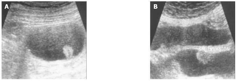
Figure 3 Images of HUS not corresponding with their histological stage.
A: Sigmoid colon cancer; HUS judged as PM cancer (T2), but histology revealed pericolic fat infiltrated by tumor cells (T3). B: Transverse colon cancer; HUS judged as PM cancer (T2), but histology revealed intact PM layer and tumor cells infiltrated only SM layer (T1).
- Citation: Chung HW, Chung JB, Park SW, Song SY, Kang JK, Park CI. Comparison of hydrocolonic sonograpy accuracy in preoperative staging between colon and rectal cancer. World J Gastroenterol 2004; 10(8): 1157-1161
- URL: https://www.wjgnet.com/1007-9327/full/v10/i8/1157.htm
- DOI: https://dx.doi.org/10.3748/wjg.v10.i8.1157


