Copyright
©The Author(s) 2004.
World J Gastroenterol. Apr 15, 2004; 10(8): 1157-1161
Published online Apr 15, 2004. doi: 10.3748/wjg.v10.i8.1157
Published online Apr 15, 2004. doi: 10.3748/wjg.v10.i8.1157
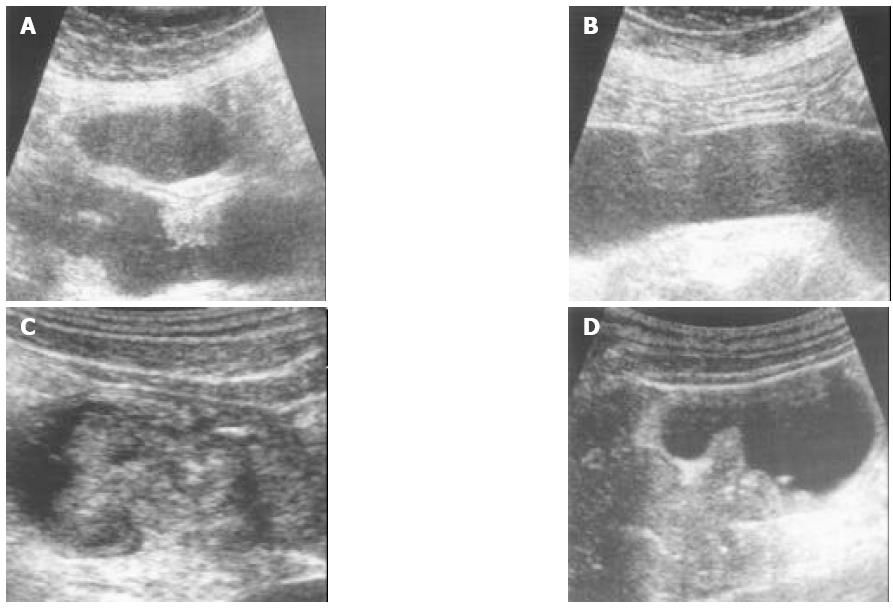
Figure 2 Images of HUS corresponding with their histological stage A: A round mass originated from submucosal (SM) layer in the descending colon, its histological stage was SM (T1).
B: A round mass with deformed PM layer (T2) in transverse colon, its histological stage was PM (T2). C: A large endoluminal polypoid mass penetrating the wall and extending to pericolic fat layer. HUS judged stage was T3, its histological stage was T3. D: A carcinoma with serosal infiltration extending into pericolic adjacent tissue in descending colon. HUS judged stage was T4, its histological stage was T4.
- Citation: Chung HW, Chung JB, Park SW, Song SY, Kang JK, Park CI. Comparison of hydrocolonic sonograpy accuracy in preoperative staging between colon and rectal cancer. World J Gastroenterol 2004; 10(8): 1157-1161
- URL: https://www.wjgnet.com/1007-9327/full/v10/i8/1157.htm
- DOI: https://dx.doi.org/10.3748/wjg.v10.i8.1157


