Copyright
©The Author(s) 2004.
World J Gastroenterol. Apr 15, 2004; 10(8): 1103-1109
Published online Apr 15, 2004. doi: 10.3748/wjg.v10.i8.1103
Published online Apr 15, 2004. doi: 10.3748/wjg.v10.i8.1103
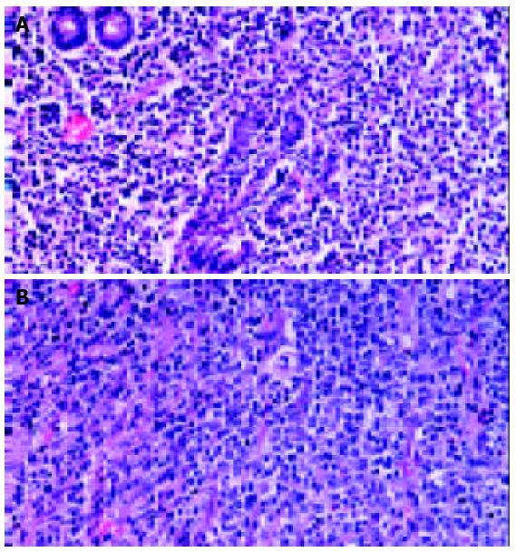
Figure 1 A: Low grade MALT lymphoma.
Microphotograph showing proliferation of heterogeneous small B-cells, includ-ing marginal zone (centrocyte-like) cells, cells resembling monocytoid cells, small lymphocytes, and scattered immunoblast and centroblast-like cells. In epithelial tissue, neo-plastic cells typically infiltrated the epithelium, forming lymphoepithelial lesions. B: Diffuse large B cell lymphoma. Microphotograph showing proliferation of large B lymphoid cells in a diffuse pattern. Tumor cells have a large and pleo-morphic appearance with prominent nucleoli.
- Citation: Kong SH, Kim MA, Park DJ, Lee HJ, Lee HS, Kim CW, Yang HK, Heo DS, Lee KU, Choe KJ. Clinicopathologic features of surgically resected primary gastric lymphoma. World J Gastroenterol 2004; 10(8): 1103-1109
- URL: https://www.wjgnet.com/1007-9327/full/v10/i8/1103.htm
- DOI: https://dx.doi.org/10.3748/wjg.v10.i8.1103


