Copyright
©The Author(s) 2004.
World J Gastroenterol. Mar 15, 2004; 10(6): 864-870
Published online Mar 15, 2004. doi: 10.3748/wjg.v10.i6.864
Published online Mar 15, 2004. doi: 10.3748/wjg.v10.i6.864
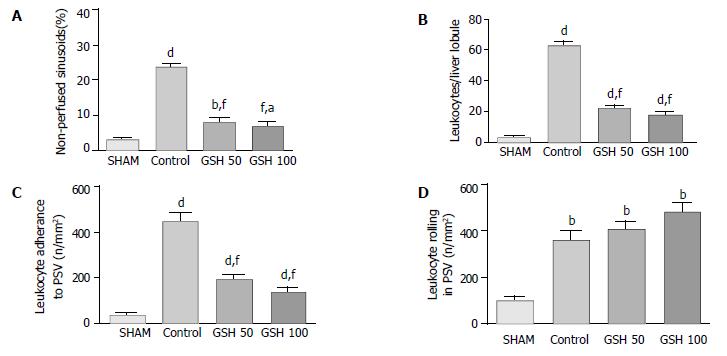
Figure 4 Impact of GSH treatment on the hepatic microcirculation after liver transplantation.
Data from in vivo microscopy were obtained 30 min after starting reperfusion comparing untreated controls (n = 8) with GSH- treated allografts (50 µmol/(h·kg) and 100 µmol/(h·kg); each n = 5) as well as to a sham group (n = 5). (A) Acinar perfusion failure is indicated as the percentage of non-perfused sinusoids (no reflow). (B) Leukocyte-adhesion (sticking) to sinusoids and (C) to postsinusoidal venules. (D) Temporary attachment of leukocytes (rolling) to the endothelium of postsinusoidal venules (PSV). Results are mean ± SE. aP < 0.05, dP < 0.01 and bP < 0.001 vs sham group. fP < 0.001 vs control group.
- Citation: Schauer RJ, Kalmuk S, Gerbes AL, Leiderer R, Meissner H, Schildberg FW, Messmer K, Bilzer M. Intravenous administration of glutathione protects parenchymal and non-parenchymal liver cells against reperfusion injury following rat liver transplantation. World J Gastroenterol 2004; 10(6): 864-870
- URL: https://www.wjgnet.com/1007-9327/full/v10/i6/864.htm
- DOI: https://dx.doi.org/10.3748/wjg.v10.i6.864


