Copyright
©The Author(s) 2004.
World J Gastroenterol. Feb 15, 2004; 10(4): 525-530
Published online Feb 15, 2004. doi: 10.3748/wjg.v10.i4.525
Published online Feb 15, 2004. doi: 10.3748/wjg.v10.i4.525
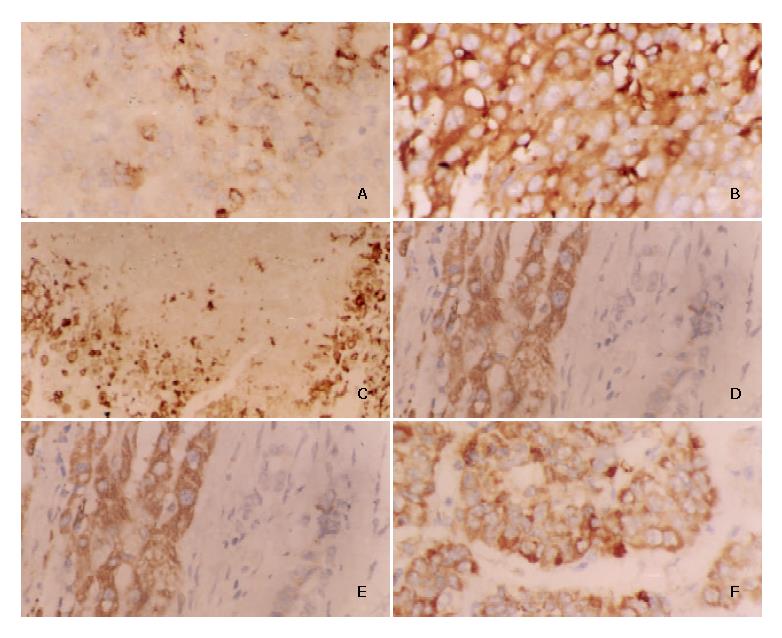
Figure 1 Characterization of HIF-2α/EPAS1 expression in human hepatocellular carcinoma tissues by IHC technique.
A: Weak expression of HIF-2α/EPAS1 in membranes and cytoplasms of HCC cells (× 400). B: Strong cytoplasmic immunoreactivity of HIF-2α/EPAS1 in HCC cells (× 400). C: HIF-2α/EPAS1 positive staining (arrows) in perinecrotic region near tumor in a HCC sample (with capsular infiltration and portal vein invasion) (× 400). D: Strong HIF-2α/EPAS1 expression in HCC tissues whereas no staining in adjacent stroma cells (× 400). E: Strong staining in the cytoplasm of macrophages compared with cancer cells, showing weak staining for HIF-2α/EPAS1 (× 400). F: Moderate to strong positive staining of HIF-2α/EPAS1 in tumor clusters infiltrating to the tissue (× 400).
- Citation: Bangoura G, Yang LY, Huang GW, Wang W. Expression of HIF-2α/EPAS1 in hepatocellular carcinoma. World J Gastroenterol 2004; 10(4): 525-530
- URL: https://www.wjgnet.com/1007-9327/full/v10/i4/525.htm
- DOI: https://dx.doi.org/10.3748/wjg.v10.i4.525


