Copyright
©The Author(s) 2004.
World J Gastroenterol. Dec 15, 2004; 10(24): 3608-3611
Published online Dec 15, 2004. doi: 10.3748/wjg.v10.i24.3608
Published online Dec 15, 2004. doi: 10.3748/wjg.v10.i24.3608
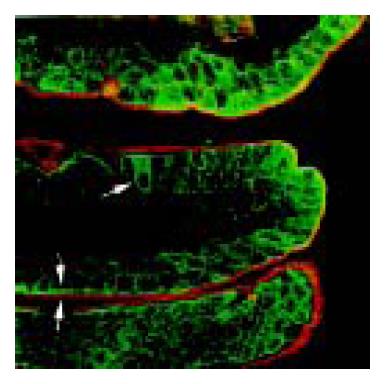
Figure 4 Double staining of immunofluorescent GAD65 (green in color) and fluorescent WGA (red in color) in rat jejunum.
Arrow points to the GAD65 strongly positive cells showing WGA negative staining. Arrowhead indicates the strong line-like staining of GAD65 along the brush border, and the outer mucus layer stained by WGA. × 630.
- Citation: Wang FY, Watanabe M, Zhu RM, Maemura K. Characteristic expression of γ-aminobutyric acid and glutamate decarboxylase in rat jejunum and its relation to differentiation of epithelial cells. World J Gastroenterol 2004; 10(24): 3608-3611
- URL: https://www.wjgnet.com/1007-9327/full/v10/i24/3608.htm
- DOI: https://dx.doi.org/10.3748/wjg.v10.i24.3608


