Copyright
©The Author(s) 2004.
World J Gastroenterol. Dec 15, 2004; 10(24): 3597-3601
Published online Dec 15, 2004. doi: 10.3748/wjg.v10.i24.3597
Published online Dec 15, 2004. doi: 10.3748/wjg.v10.i24.3597
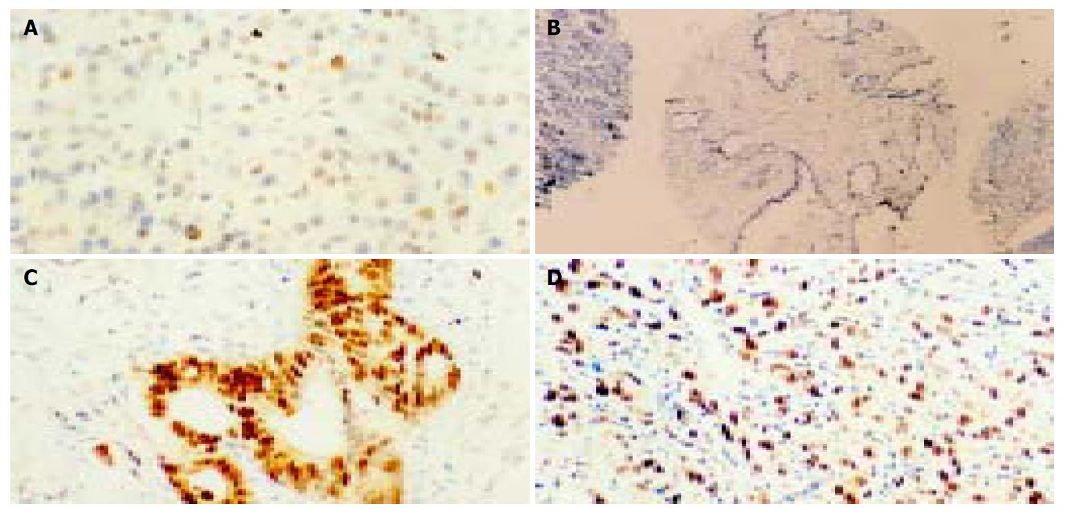
Figure 1 Representative examples of IHC staining for p33ING1b (× 200) on normal pancreatic tissues and cancer tissues.
A: Weak positive pancreatic cells for p33ING1b; B: No p33ING1b; C and D: Extremely strong staining in pancreatic cancer samples.
- Citation: Yu GZ, Zhu MH, Zhu Z, Ni CR, Zheng JM, Li FM. Genetic alterations and reduced expression of tumor suppressor p33ING1b in human exocrine pancreatic carcinoma. World J Gastroenterol 2004; 10(24): 3597-3601
- URL: https://www.wjgnet.com/1007-9327/full/v10/i24/3597.htm
- DOI: https://dx.doi.org/10.3748/wjg.v10.i24.3597


