Copyright
©The Author(s) 2004.
World J Gastroenterol. Jan 15, 2004; 10(2): 238-243
Published online Jan 15, 2004. doi: 10.3748/wjg.v10.i2.238
Published online Jan 15, 2004. doi: 10.3748/wjg.v10.i2.238
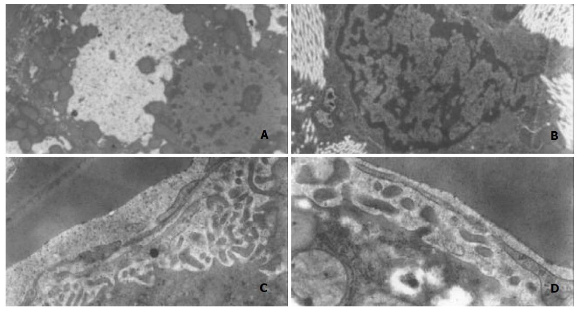
Figure 3 Changes of hepatocytes observed by electron microscopy.
A: fatty vesicle in hepatocytes, B: activated HSC and fibril, C: sinusoidal endothelium and basement, D: endothelium,less fenestrae.
- Citation: Xu GF, Wang XY, Ge GL, Li PT, Jia X, Tian DL, Jiang LD, Yang JX. Dynamic changes of capillarization and peri-sinusoid fibrosis in alcoholic liver diseases. World J Gastroenterol 2004; 10(2): 238-243
- URL: https://www.wjgnet.com/1007-9327/full/v10/i2/238.htm
- DOI: https://dx.doi.org/10.3748/wjg.v10.i2.238


