Copyright
©The Author(s) 2004.
World J Gastroenterol. Aug 15, 2004; 10(16): 2417-2418
Published online Aug 15, 2004. doi: 10.3748/wjg.v10.i16.2417
Published online Aug 15, 2004. doi: 10.3748/wjg.v10.i16.2417
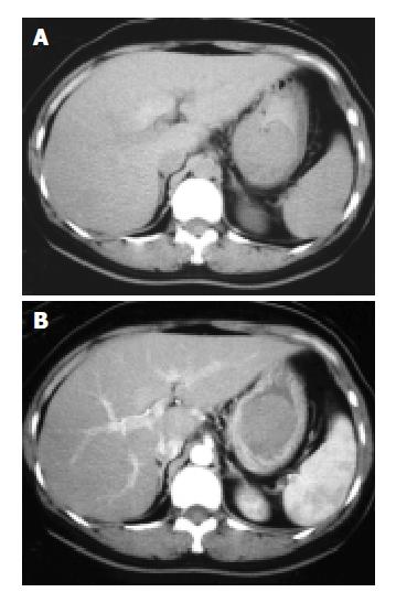
Figure 1 Precontrast and postcontrast CT scans of tumor.
A: A precontrast CT scan shows a well-defined intragastric tumor with slightly lower density than the liver. The hounsfield units are 38.77. B: A postcontrast CT scan shows homogenous en-hancement of the tumor, with a hounsfield unit of 57.58.
- Citation: Lee CM, Chen HC, Leung TK, Chen YY. Gastrointestinal stromal tumor: Computed tomographic features. World J Gastroenterol 2004; 10(16): 2417-2418
- URL: https://www.wjgnet.com/1007-9327/full/v10/i16/2417.htm
- DOI: https://dx.doi.org/10.3748/wjg.v10.i16.2417


