Copyright
©The Author(s) 2004.
World J Gastroenterol. Aug 15, 2004; 10(16): 2373-2378
Published online Aug 15, 2004. doi: 10.3748/wjg.v10.i16.2373
Published online Aug 15, 2004. doi: 10.3748/wjg.v10.i16.2373
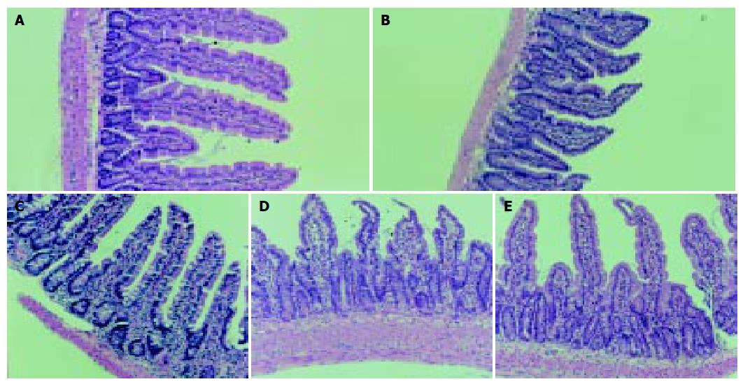
Figure 1 Alterations in architecture of small intestinal mucosa.
(HE staining, original magnification: × 100). A: Normal mucous membrane of jejunum in control rats; B: Normal mucous membrane of ileum in control rats; C: Histological changes of jejunal mucosa of group T1 rats. The villi become sparser than those in control. The height and shape of villi are normal on the whole; D: Histological changes of ileal mucosa in PN group rats. The section shows a severe atrophy in mucosal structure. The villi become shorter, blunt and swollen; E: Histological changes of jejunal mucosa of Sep group. The villi are sparse and shorter than those of control group.
- Citation: Ding LA, Li JS, Li YS, Zhu NT, Liu FN, Tan L. Intestinal barrier damage caused by trauma and lipopolysaccharide. World J Gastroenterol 2004; 10(16): 2373-2378
- URL: https://www.wjgnet.com/1007-9327/full/v10/i16/2373.htm
- DOI: https://dx.doi.org/10.3748/wjg.v10.i16.2373


