Copyright
©The Author(s) 2004.
World J Gastroenterol. Aug 15, 2004; 10(16): 2344-2351
Published online Aug 15, 2004. doi: 10.3748/wjg.v10.i16.2344
Published online Aug 15, 2004. doi: 10.3748/wjg.v10.i16.2344
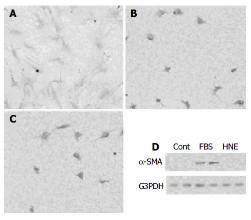
Figure 9 A,B,C: Freshly isolated PSCs were incubated with 50 mL/L serum (panel A), HNE (at 1 µmol/L, panel B), or serum-free medium (panel C) for 7 d.
Morphological changes characteristic of PSC activation were assessed after staining with GFAP. Original magnification: × 10 objective. D: Total cell lysates (approximately 25 µg) were prepared from cells treated with serum-free medium (“Cont”), 50 mL/L serum (“FBS”), or HNE (at 1 µmol/L) for 7 d, and the levels of α -SMA and G3PDH were determined by Western blotting.
- Citation: Kikuta K, Masamune A, Satoh M, Suzuki N, Shimosegawa T. 4-hydroxy-2, 3-nonenal activates activator protein-1 and mitogen-activated protein kinases in rat pancreatic stellate cells. World J Gastroenterol 2004; 10(16): 2344-2351
- URL: https://www.wjgnet.com/1007-9327/full/v10/i16/2344.htm
- DOI: https://dx.doi.org/10.3748/wjg.v10.i16.2344


