Copyright
©The Author(s) 2004.
World J Gastroenterol. Aug 1, 2004; 10(15): 2287-2291
Published online Aug 1, 2004. doi: 10.3748/wjg.v10.i15.2287
Published online Aug 1, 2004. doi: 10.3748/wjg.v10.i15.2287
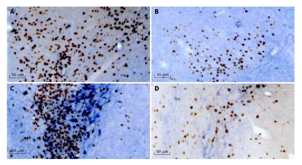
Figure 1 Photomicrographs showing c-Fos and NOS positive neurons in amygdale (A and B), paraventricular nucleus (C and D).
A and C were taken from rats with acid-pepsin perfusion, while B and D were taken from rats with saline perfusion. (3V: the third ventricle).
- Citation: Shuai XW, Xie PY. Expression and localization of c-Fos and NOS in the central nerve system following esophageal acid stimulation in rats. World J Gastroenterol 2004; 10(15): 2287-2291
- URL: https://www.wjgnet.com/1007-9327/full/v10/i15/2287.htm
- DOI: https://dx.doi.org/10.3748/wjg.v10.i15.2287


