Copyright
©The Author(s) 2004.
World J Gastroenterol. Aug 1, 2004; 10(15): 2201-2204
Published online Aug 1, 2004. doi: 10.3748/wjg.v10.i15.2201
Published online Aug 1, 2004. doi: 10.3748/wjg.v10.i15.2201
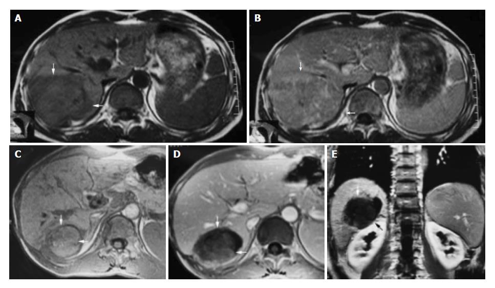
Figure 1 A 42-year-old male with hepatocellular carcinoma.
A: MRI T1-weighted images before HIFU treatment showed that liver tumor of right-posterior lobe was 115 mm × 100 mm × 66 mm; B: MRI enhanced imaging before HIFU revealed that enhancement of mass in liver right-posterior lobe was higher than that of surrounding normal liver tissues; C: MRI T1-weighted imaging 11 mo after HIFU revealed that liver tumor of right-posterior lobe obviously decreased in size (50 mm × 55 mm × 60 mm); D, E: MRI enhanced imaging (cross section and coronal section) 11 mo after HIFU revealed that liver tumor had shrunk obviously, blood supply of tumor was eliminated, the tumor therapeutic region had coagulation necrosis.
- Citation: Li CX, Xu GL, Jiang ZY, Li JJ, Luo GY, Shan HB, Zhang R, Li Y. Analysis of clinical effect of high-intensity focused ultrasound on liver cancer. World J Gastroenterol 2004; 10(15): 2201-2204
- URL: https://www.wjgnet.com/1007-9327/full/v10/i15/2201.htm
- DOI: https://dx.doi.org/10.3748/wjg.v10.i15.2201


