Copyright
©The Author(s) 2004.
World J Gastroenterol. Aug 1, 2004; 10(15): 2179-2183
Published online Aug 1, 2004. doi: 10.3748/wjg.v10.i15.2179
Published online Aug 1, 2004. doi: 10.3748/wjg.v10.i15.2179
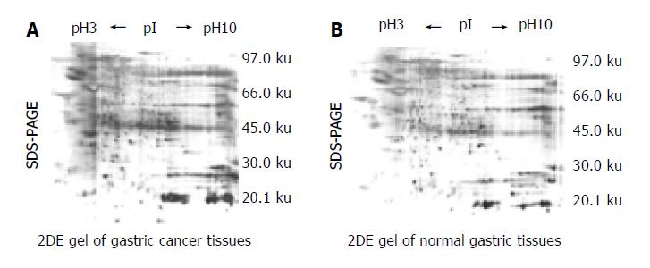
Figure 1 2DE maps of human gastric tissue from No.
5 patient. A: was from gastric cancer. B: from normal gastric sample of the same patient. Proteins were separated on pH 3 -10 linear IPG strip in the first dimension and 125 g/L SDS-PAGE in the second dimension, gels were silver stained. All labeled spots were tumor-specific.
- Citation: Wang KJ, Wang RT, Zhang JZ. Identification of tumor markers using two-dimensional electrophoresis in gastric carcinoma. World J Gastroenterol 2004; 10(15): 2179-2183
- URL: https://www.wjgnet.com/1007-9327/full/v10/i15/2179.htm
- DOI: https://dx.doi.org/10.3748/wjg.v10.i15.2179


