Copyright
©The Author(s) 2004.
World J Gastroenterol. Jul 15, 2004; 10(14): 2003-2009
Published online Jul 15, 2004. doi: 10.3748/wjg.v10.i14.2003
Published online Jul 15, 2004. doi: 10.3748/wjg.v10.i14.2003
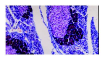
Figure 1 Histologic alterations in the pancreas following Arg-induced pancreatitis on d 3.
The signs of acute inflammation and tubular complexes are visible. Segmental fibroses with blue stained fibroblast are observed. Intact pancreatic acinar cells are presented mainly around the Langerhans islets (Chrossmonn’s trichrome staining × 100).
- Citation: Hegyi P, Jr ZR, Sári R, Góg C, Lonovics J, Takács T, Czakó L. L-arginine-induced experimental pancreatitis. World J Gastroenterol 2004; 10(14): 2003-2009
- URL: https://www.wjgnet.com/1007-9327/full/v10/i14/2003.htm
- DOI: https://dx.doi.org/10.3748/wjg.v10.i14.2003


