Copyright
©The Author(s) 2004.
World J Gastroenterol. Jul 1, 2004; 10(13): 1943-1947
Published online Jul 1, 2004. doi: 10.3748/wjg.v10.i13.1943
Published online Jul 1, 2004. doi: 10.3748/wjg.v10.i13.1943
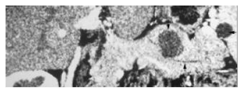
Figure 4 Neuroendocrine tumor of pancreas and peripancreatic pseudocyst.
Curved plane showed a non-enhanced hypoattenuation mass located in the pancreatic parenchyma (arrow) and a pseudocyst at the port of spleen during the pan-creatic parenchyma phase. The normal pancreatic duct was also well depicted (long arrow).
- Citation: Gong JS, Xu JM. Role of curved planar reformations using multidetector spiral CT in diagnosis of pancreatic and peripancreatic diseases. World J Gastroenterol 2004; 10(13): 1943-1947
- URL: https://www.wjgnet.com/1007-9327/full/v10/i13/1943.htm
- DOI: https://dx.doi.org/10.3748/wjg.v10.i13.1943


