Copyright
©The Author(s) 2004.
World J Gastroenterol. Jun 1, 2004; 10(11): 1594-1599
Published online Jun 1, 2004. doi: 10.3748/wjg.v10.i11.1594
Published online Jun 1, 2004. doi: 10.3748/wjg.v10.i11.1594
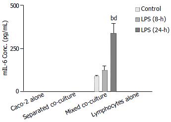
Figure 7 Comparison of Shigella F2a-12 LPS-induced mIL-6 release from different culture groups by ELISA.
Culture groups and Peyer’s patch lymphocytes (1 × 10⁶ cells) were treated with LPS for 8-h and 24-h. The values indicate mean ± SE; n = 5; bP < 0.01 (compared to mixed co-culture control) and dP < 0.01 (compared to the mixed LPS treatment for 8-h).
- Citation: Chen J, Tsang LL, Ho LS, Rowlands DK, Gao JY, Ng CP, Chung YW, Chan HC. Modulation of human enteric epithelial barrier and ion transport function by Peyer’s patch lymphocytes. World J Gastroenterol 2004; 10(11): 1594-1599
- URL: https://www.wjgnet.com/1007-9327/full/v10/i11/1594.htm
- DOI: https://dx.doi.org/10.3748/wjg.v10.i11.1594


