Copyright
©The Author(s) 2004.
World J Gastroenterol. May 15, 2004; 10(10): 1480-1486
Published online May 15, 2004. doi: 10.3748/wjg.v10.i10.1480
Published online May 15, 2004. doi: 10.3748/wjg.v10.i10.1480
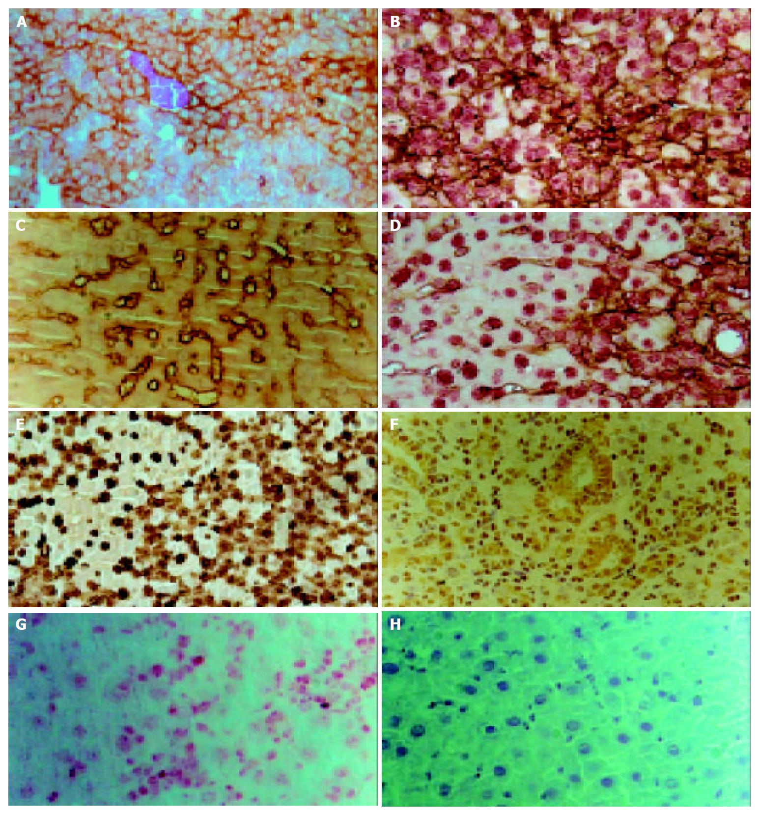
Figure 3 Immunohistochemical staining of liver sections from rat liver exposed to 2-AAF/PH (d 11) at high magnification.
A: Staining for OV6, Arrows point to OV6-positive oval cells.B: Double immunohitstochemical staining for OV6 (brown) and albu-min (red). C: Staining for cytokeratin19 (CK19). Ductular lumen face and oval cells were positive. Arrows point ot CK19-positive cells. D: Double immunhistochemical staining for CK19 (brown) and albumin (red). Arrows point to oval cells with tow markers. E: Staining for AFP. F: Staining for connexin43, Arrows point to connexin43 positive oval cells. G: Staining for C-kit, Oval cells were stained with red. H: Negative control.
- Citation: Qin AL, Zhou XQ, Zhang W, Yu H, Xie Q. Characterization and enrichment of hepatic progenitor cells in adult rat liver. World J Gastroenterol 2004; 10(10): 1480-1486
- URL: https://www.wjgnet.com/1007-9327/full/v10/i10/1480.htm
- DOI: https://dx.doi.org/10.3748/wjg.v10.i10.1480


