Copyright
©The Author(s) 2004.
World J Gastroenterol. May 15, 2004; 10(10): 1480-1486
Published online May 15, 2004. doi: 10.3748/wjg.v10.i10.1480
Published online May 15, 2004. doi: 10.3748/wjg.v10.i10.1480
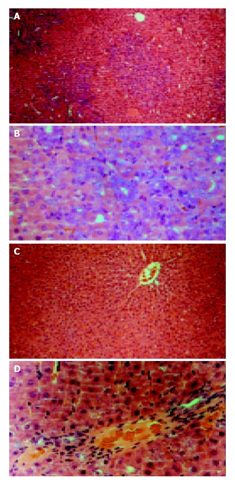
Figure 1 Rat liver sections obtained from 2-AAF/PH (11 d after operation) and control.
Sections were stained with he-matoxylin-eosin. (A and B) show liver sections obtained from 2-AAF/PH-treated rats at low (× 100) and high (× 400) magnification. Small oval cells (arrows) can be seen close prox-imity to proliferating bile ducts and in areas of ductular pro-liferation or in acinar arrangements around hepatocytes. The oval cells radiate from the periportal region, forming primi-tive ductular structures with poorly defined lumen. (C and D) show liver tissue from control rats at low and high magnification. Hepatoctye proliferation can be seen in the typical liver architecture. Central vein (CV) and portal triad region can be seen.
- Citation: Qin AL, Zhou XQ, Zhang W, Yu H, Xie Q. Characterization and enrichment of hepatic progenitor cells in adult rat liver. World J Gastroenterol 2004; 10(10): 1480-1486
- URL: https://www.wjgnet.com/1007-9327/full/v10/i10/1480.htm
- DOI: https://dx.doi.org/10.3748/wjg.v10.i10.1480


