Copyright
©The Author(s) 2004.
World J Gastroenterol. May 15, 2004; 10(10): 1471-1475
Published online May 15, 2004. doi: 10.3748/wjg.v10.i10.1471
Published online May 15, 2004. doi: 10.3748/wjg.v10.i10.1471
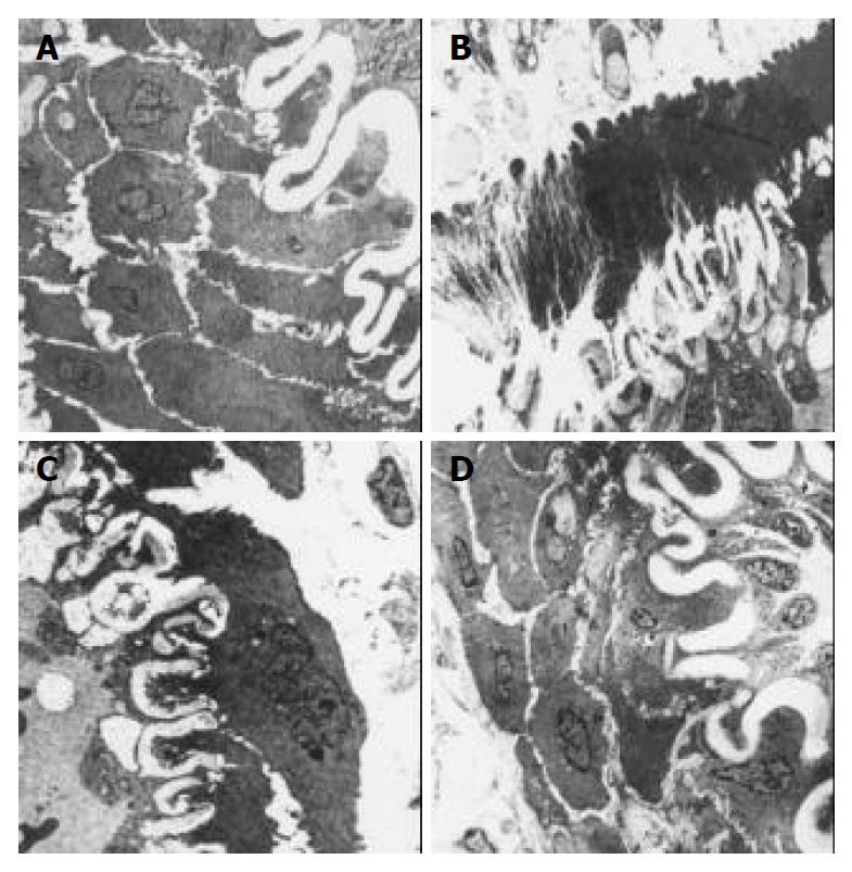
Figure 2 Microstructure of mesenteric arteries (transmission electron microscope), A: WKY group, endothelial cells of in-tima were abundant and normal, Media has more smooth muscle cells, internal elastic lamina is normal (× 2000).
B: SHR group, endothelial cells of intima had vacuole and fibrous tis-sue with adventitial hyperplasia; thickness of the adventitia was increased; media was severely fibrous and the fibrous tissue extended to smooth muscle layer and invaded internal elastic lamina; internal elastic lamina was tortuous and atrophic; some of smooth muscle cells were replaced by fibrous tissue (× 2500). C: Irbesartan group, endothelial cells of intima have vacuole and marrow type corpses, internal elastic lamina was tortuous and atrophic and infiltrated by collagen fibers (× 2500). D: Imidapril group, endothelial cells of intima have marrow type corpses but no vacuole, numbers of smooth muscle cells in media were more than those in irbesartan group; internal elastic lamina got a close-to-normal distribution; local internal elastic lamina got narrow, fibrosis did not occur on blood ves-sel wall (× 2000).
- Citation: Zhu ZS, Wang JM, Chen SL. Mesenteric artery remodeling and effects of imidapril and irbesartan on it in spontaneously hypertensive rats. World J Gastroenterol 2004; 10(10): 1471-1475
- URL: https://www.wjgnet.com/1007-9327/full/v10/i10/1471.htm
- DOI: https://dx.doi.org/10.3748/wjg.v10.i10.1471


