Copyright
©The Author(s) 2004.
World J Gastroenterol. May 15, 2004; 10(10): 1415-1420
Published online May 15, 2004. doi: 10.3748/wjg.v10.i10.1415
Published online May 15, 2004. doi: 10.3748/wjg.v10.i10.1415
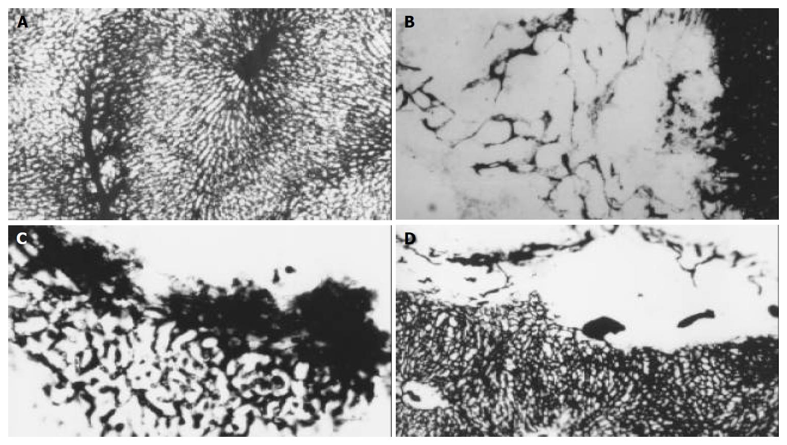
Figure 3 Micro-vessel casting with Chinese ink through ascending artery.
A: The specimens show a lobular architecture of normal liver, magnification × 100. B: In control group, hepatic artery perfusion shows networks of micro-vessels or plexuses of dilated and tortuous course around and within the tumor originated from the arterioles, some sinusoid vessels can be observed in this tumor, magnification × 100. C: The original micro-vessels of the tumor are remarkably diminished, small and dense new plexuses appear around the tumor in lipiodol group, which are correlated to intense reaction of granulation tissue, magnification × 100. D: micro-vessels are decreased in bletilla group, no new micro-vessels can be seen at all, magnification × 100.
- Citation: Zhao JG, Feng GS, Kong XQ, Li X, Li MH, Cheng YS. Changes of tumor microcirculation after transcatheter arterial chemoembolization: First pass perfusion MR imaging and Chinese ink casting in a rabbit model. World J Gastroenterol 2004; 10(10): 1415-1420
- URL: https://www.wjgnet.com/1007-9327/full/v10/i10/1415.htm
- DOI: https://dx.doi.org/10.3748/wjg.v10.i10.1415


