Copyright
©The Author(s) 2004.
World J Gastroenterol. May 15, 2004; 10(10): 1415-1420
Published online May 15, 2004. doi: 10.3748/wjg.v10.i10.1415
Published online May 15, 2004. doi: 10.3748/wjg.v10.i10.1415
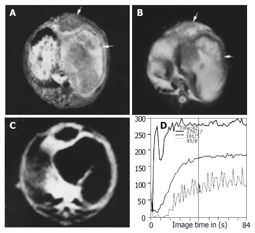
Figure 2 Images obtained after treatment in lipiodol group.
Inhomogeneous hyperintense lesions with intermediate intense rim (arrow head) can be seen on T1WI (A) and T2WI (B), indicating the necrosis of the lesion and intratumor retention of lipiodol. (C) FP T1-weighted image with gadolinium shows thinner rim enhancement and no enhancement in the center of the lesion com-pared with control group although the lesion volume is increased. (D) SS of the curve in center of the lesion is decreased compared with those of the border.
- Citation: Zhao JG, Feng GS, Kong XQ, Li X, Li MH, Cheng YS. Changes of tumor microcirculation after transcatheter arterial chemoembolization: First pass perfusion MR imaging and Chinese ink casting in a rabbit model. World J Gastroenterol 2004; 10(10): 1415-1420
- URL: https://www.wjgnet.com/1007-9327/full/v10/i10/1415.htm
- DOI: https://dx.doi.org/10.3748/wjg.v10.i10.1415


