Copyright
©The Author(s) 2004.
World J Gastroenterol. May 15, 2004; 10(10): 1387-1391
Published online May 15, 2004. doi: 10.3748/wjg.v10.i10.1387
Published online May 15, 2004. doi: 10.3748/wjg.v10.i10.1387
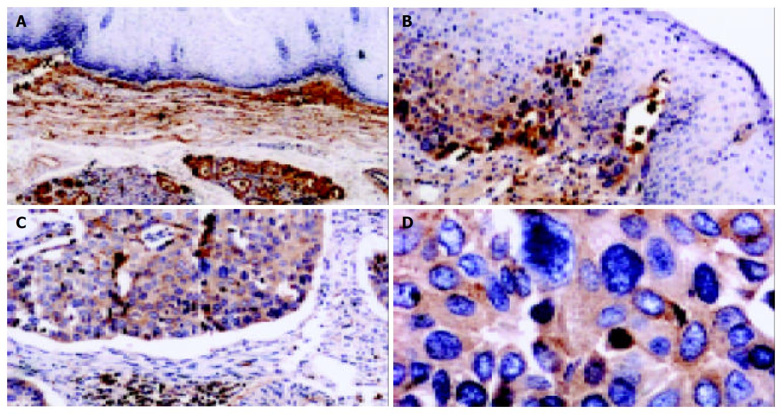
Figure 4 Detection of clusterin protein in tissue sections of human ESCC by immunohistochemistry.
In normal esophageal mucosa, the expression of clusterin in squamous epithelial cells and basal lamina was negative, while clusterin immunostaining was visualized obviously in the stroma of epithelial mucosa (A, 100 ×); clusterin was positive in middle dysplasia, while the vast majority of stromal cells were negative in normal squamous epithelia of esophagus (B, 100 ×); clusterin was strong positive in the remnants of stromal extracellular matrix invaded by tumor cells in well-differentiated ESCC (C, 100 ×). D show that stromal and basal membrane are almost completely disrupted and invaded by tumor cells. (D, same section, different field of that of C, 400 ×). Counterstaining was performed with hematoxylin.
- Citation: He HZ, Song ZM, Wang K, Teng LH, Liu F, Mao YS, Lu N, Zhang SZ, Wu M, Zhao XH. Alterations in expression, proteolysis and intracellular localizations of clusterin in esophageal squamous cell carcinoma. World J Gastroenterol 2004; 10(10): 1387-1391
- URL: https://www.wjgnet.com/1007-9327/full/v10/i10/1387.htm
- DOI: https://dx.doi.org/10.3748/wjg.v10.i10.1387


