Copyright
©The Author(s) 2004.
World J Gastroenterol. May 15, 2004; 10(10): 1387-1391
Published online May 15, 2004. doi: 10.3748/wjg.v10.i10.1387
Published online May 15, 2004. doi: 10.3748/wjg.v10.i10.1387
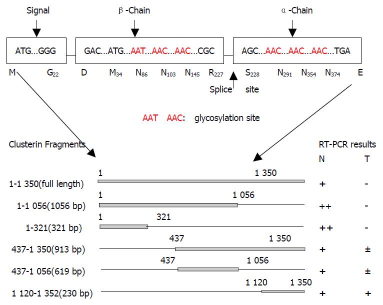
Figure 1 Schematic diagram of analyzed fragments of clusterin.
A: structural map of the human clusterin protein. Letters and boxes signify the following: ER-targeting signal, endoplasmic reticulum-targeting hydrophobic leader peptide; splice site, α/β-cleavage site (amino acid 205 and 206) of cytoplasmic - 60 ku pre-secreted clusterin which was glycosylated and cleavaged into α and β chains to form an 80 ku matured secre-tory clusterin protein; rectangles, hydrophobic leader peptide (N-terminal 66 bp encode aa M1 to G22, 22 aa), β-chain (5’ end) and α-chain (3’ end); Red letters, the glycosylation sites. B: Schematic diagram of analyzed fragments of clusterin and their RT-PCR results.
- Citation: He HZ, Song ZM, Wang K, Teng LH, Liu F, Mao YS, Lu N, Zhang SZ, Wu M, Zhao XH. Alterations in expression, proteolysis and intracellular localizations of clusterin in esophageal squamous cell carcinoma. World J Gastroenterol 2004; 10(10): 1387-1391
- URL: https://www.wjgnet.com/1007-9327/full/v10/i10/1387.htm
- DOI: https://dx.doi.org/10.3748/wjg.v10.i10.1387


