Copyright
©The Author(s) 2004.
World J Gastroenterol. Jan 1, 2004; 10(1): 67-72
Published online Jan 1, 2004. doi: 10.3748/wjg.v10.i1.67
Published online Jan 1, 2004. doi: 10.3748/wjg.v10.i1.67
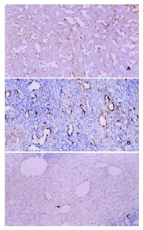
Figure 3 Three patterns of intratumoral MVD distribution revealed by F8RA immunohistochemical staining.
A: Pattern I. Markedly dilated and abundant blood sinusoids with very rich positively stained sinusoidal endothelial cells and scanty tumor interstitium. B: Pattern II. Sinusoids and interstitium abundant and rich in positively stained endothelial cells (black arrow). C: Pattern III. Rich tumor interstitium with few posi-tively stained endothelial cells (white arrow) and scanty blood sinusoids.
- Citation: Chen WX, Min PQ, Song B, Xiao BL, Liu Y, Ge YH. Single-level dynamic spiral CT of hepatocellular carcinoma: Correlation between imaging features and density of tumor microvessels. World J Gastroenterol 2004; 10(1): 67-72
- URL: https://www.wjgnet.com/1007-9327/full/v10/i1/67.htm
- DOI: https://dx.doi.org/10.3748/wjg.v10.i1.67


