Copyright
©The Author(s) 2004.
World J Gastroenterol. Jan 1, 2004; 10(1): 67-72
Published online Jan 1, 2004. doi: 10.3748/wjg.v10.i1.67
Published online Jan 1, 2004. doi: 10.3748/wjg.v10.i1.67
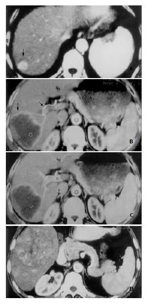
Figure 2 Three types of enhancement morphology depicted in HCC patients.
A: Type A. Marked and homogeneous enhance-ment of the entire HCC lesion in the posterosuperior segment of right hepatic lobe (black arrow). B: Type B. Bright periph-eral ring-like enhancement of HCC lesion on arterial phase image in the right posterosuperior segment (black arrow), and the dilated right hepatic artery (black arrow). C: Portal venous image at the same slice level as in (B). HCC lesion remained hypodense despite obvious enhancement of normal liver pa-renchyma elsewhere. D: Type C. Inhomogeneous patchy en-hancement of HCC lesion in right lower hepatic lobe from ar-terial phase image. Bright dots and linear shadows represent enhanced tumor vessels within the lesion.
- Citation: Chen WX, Min PQ, Song B, Xiao BL, Liu Y, Ge YH. Single-level dynamic spiral CT of hepatocellular carcinoma: Correlation between imaging features and density of tumor microvessels. World J Gastroenterol 2004; 10(1): 67-72
- URL: https://www.wjgnet.com/1007-9327/full/v10/i1/67.htm
- DOI: https://dx.doi.org/10.3748/wjg.v10.i1.67


