Copyright
©The Author(s) 2004.
World J Gastroenterol. Jan 1, 2004; 10(1): 53-57
Published online Jan 1, 2004. doi: 10.3748/wjg.v10.i1.53
Published online Jan 1, 2004. doi: 10.3748/wjg.v10.i1.53
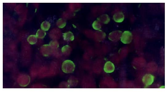
Figure 4 SEA expressed on the membrane of HepG2 (indirect immunofluorescence).
HepG2-SEA cells were stained accord-ing to the standard protocol of indirect immunofluorescence. First antibody is mouse anti-SEA IgG and second antibody is FITC labeled sheep anti-mouse IgG diluted in 0.1‰ evan blue. Positive signal is green while the background is red. SEA was located on the membrane of HepG2. Untransfected HepG2 cells did not express SEA (data not show).
- Citation: Lu SY, Sui YF, Li ZS, Ye J, Dong HL, Qu P, Zhang XM, Wang WY, Li YS. Superantigen-SEA gene modified tumor vaccine for hepatocellular carcinoma: An in vitro study. World J Gastroenterol 2004; 10(1): 53-57
- URL: https://www.wjgnet.com/1007-9327/full/v10/i1/53.htm
- DOI: https://dx.doi.org/10.3748/wjg.v10.i1.53


