Copyright
©The Author(s) 2003.
World J Gastroenterol. Apr 15, 2003; 9(4): 784-787
Published online Apr 15, 2003. doi: 10.3748/wjg.v9.i4.784
Published online Apr 15, 2003. doi: 10.3748/wjg.v9.i4.784
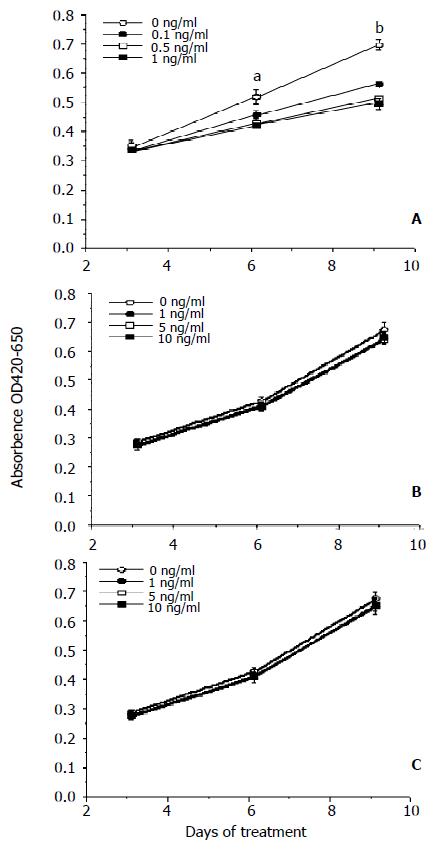
Figure 1 Regulating effect of TGF-β1, BMP-2 and BMP-4 on the proliferation of rat hepatic stellate cells.
Hepatic stellate cells were incubated with TGF-β1 (A), BMP-2 (B) and BMP-4 (C) as described in Materials and methods. The data represent mean ± SE from ten wells. The experiments were repeated two times. Denotes: a represents aP < 0.05 vs the concentrations of 0.1, 0.5 and 1 ng/mL, b means bP < 0.01vs the concentrations of 0.1, 0.5 and 1 ng/mL.
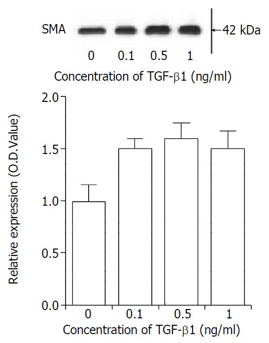
Figure 2 Regulating effect of TGF-β1 on rat hepatic stellate cell trans-differentiation.
Hepatic stellate cells were incubated with different concentrations of TGF-β1 for three days. The media and TGF-β1 were changed every other day. Western blot was performed as described in Materials and methods. The top panel represents typical Western blot of SMA. The lower panel represents histogram of densitometric data from four-separated Western blot (mean ± SE).
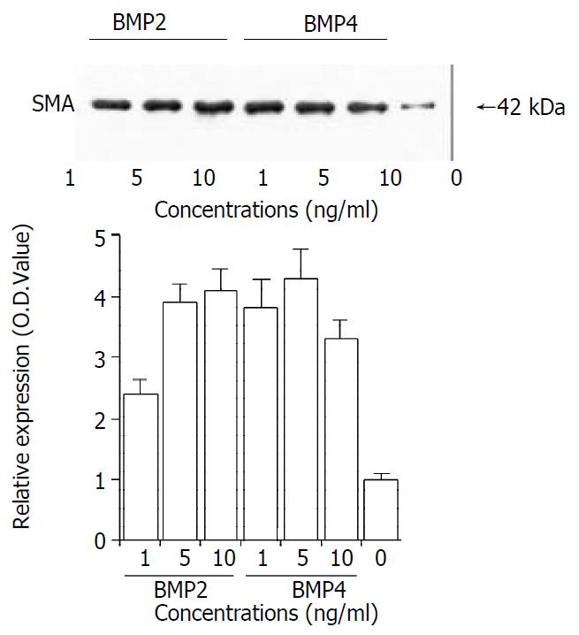
Figure 3 Regulating effect of BMP-2 and BMP-4 on hepatic stellate cell trans-differentiation.
Hepatic stellate cells were in-cubated with different concentrations of BMP-2 or BMP-4 for three days. The media and BMPs were changed every other day. Western blot was performed as described in Materials and methods. The top panel represents typical Western blot of SMA. The lower panel represents histogram of densitometric data from four-separated Western blot (mean ± SE).
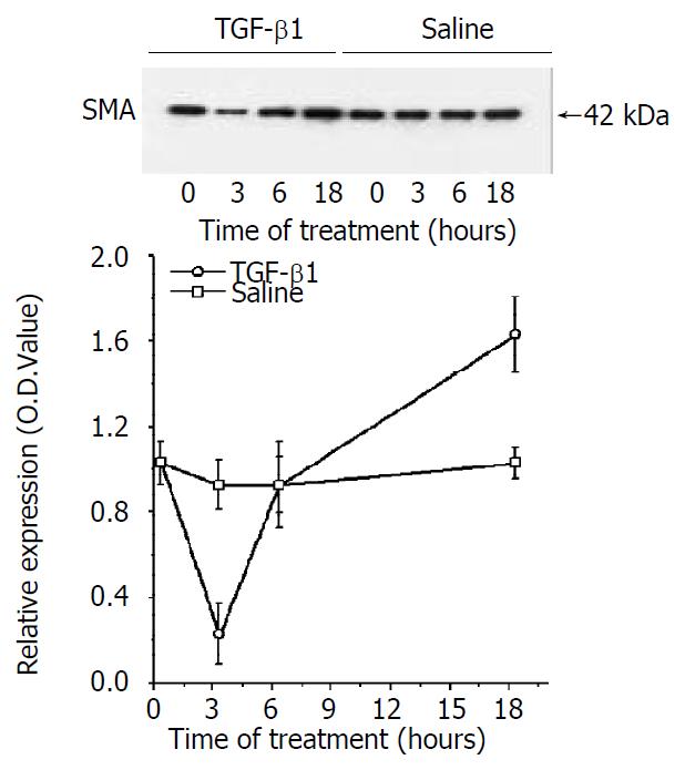
Figure 4 Regulating effect of TGF-β1 on SMA protein expression.
Hepatic stellate cells were cultured for three days and the media were removed. Media with 1 ng/mL TGF-β1 or saline were added into culture dishes and cells were collected at different times as indicated. The data represent mean ± SE from four experiments.
- Citation: Shen H, Huang GJ, Gong YW. Effect of transforming growth factor beta and bone morphogenetic proteins on rat hepatic stellate cell proliferation and trans-differentiation. World J Gastroenterol 2003; 9(4): 784-787
- URL: https://www.wjgnet.com/1007-9327/full/v9/i4/784.htm
- DOI: https://dx.doi.org/10.3748/wjg.v9.i4.784


