Copyright
©The Author(s) 2003.
World J Gastroenterol. Apr 15, 2003; 9(4): 650-654
Published online Apr 15, 2003. doi: 10.3748/wjg.v9.i4.650
Published online Apr 15, 2003. doi: 10.3748/wjg.v9.i4.650
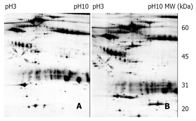
Figure 1 2-DE profiles of the pre- and post-surgery matched esophageal cancer sera from a same individual.
One hundred and eighty microgram of protein was separated by 2-DE (IEF at pH3-10, 13% of SDS-PAGE) and stained by silver staining. LMW markers were from Amersham Pharmacia. (A) pre-sur-gery serum, (B) matched post-surgery serum.
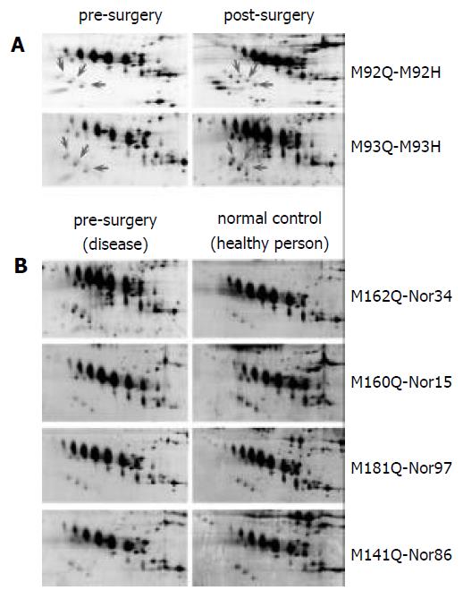
Figure 2 Representative regions of 2-DE patterns of more matched esophageal cancer sera.
One hundred and eighty microgram of protein was separated by 2-DE (IEF at pH3-10, SDS-PAGE 13%) and stained by silver staining. LMW mark-ers were from Amersham Pharmacia. (A) pre-surgery sera vs post-surgery sera, (B) pre-surgery sera vs normal control sera.
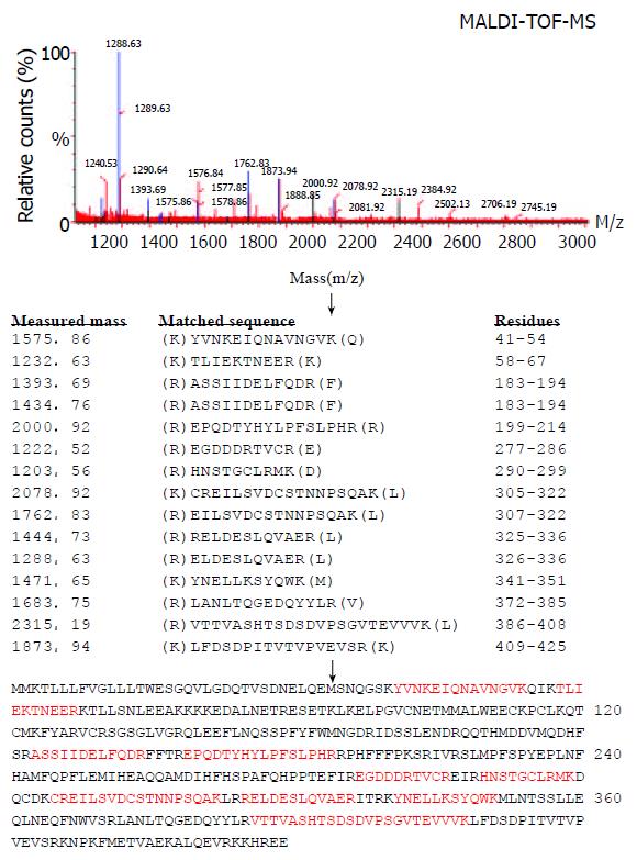
Figure 3 The spectra of MALDI-TOF-MS obtained from one of the differentiated polypeptide spots matched with the tryptic peptide sequences of clusterin (characters in red).
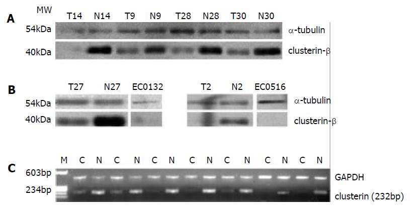
Figure 4 (A) Western blot analysis of clusterin proteins expressed in patient-matched normal and tumor epithelium; (B) Western blot analysis of clusterin proteins expressed in two kinds of esophageal squamous cell carcinoma, EC0156 and EC0132.
Alpha-tubulin was used as a loading control. (C) Expression analysis of clusterin in patient-matched esophageal cancer tissues by semi-quantitative RT-PCR. GAPDH was used as an internal control.
-
Citation: Zhang LY, Ying WT, Mao YS, He HZ, Liu Y, Wang HX, Liu F, Wang K, Zhang DC, Wang Y, Wu M, Qian XH, Zhao XH. Loss of clusterin both in serum and tissue correlates with the tumorigenesis of esophageal squamous cell carcinoma
via proteomics approaches. World J Gastroenterol 2003; 9(4): 650-654 - URL: https://www.wjgnet.com/1007-9327/full/v9/i4/650.htm
- DOI: https://dx.doi.org/10.3748/wjg.v9.i4.650


