Copyright
©The Author(s) 2003.
World J Gastroenterol. Mar 15, 2003; 9(3): 606-608
Published online Mar 15, 2003. doi: 10.3748/wjg.v9.i3.606
Published online Mar 15, 2003. doi: 10.3748/wjg.v9.i3.606
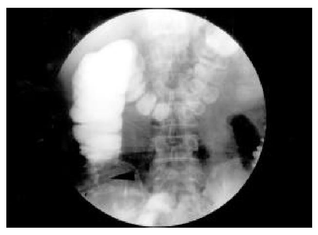
Figure 1 Barium contrast roentgenogram demonstrateed a right-sided colonic mass.
(black arrow head).
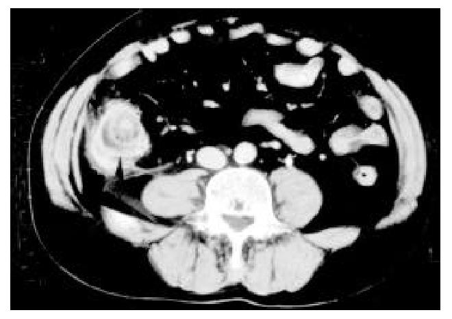
Figure 2 Acute diverticulitis of cecum in 50-year-old man.
CT scan clearly showed enhancement of thickened diverticular wall and preservation of wall enhancement pattern of cecum as hyperattenuating inner layer, thickened middle layer of low attenuation, and outer high-attenuation layer (black arrow head).
- Citation: Shyung LR, Lin SC, Shih SC, Kao CR, Chou SY. Decision making in right-sided diverticulitis. World J Gastroenterol 2003; 9(3): 606-608
- URL: https://www.wjgnet.com/1007-9327/full/v9/i3/606.htm
- DOI: https://dx.doi.org/10.3748/wjg.v9.i3.606


