Copyright
©The Author(s) 2003.
World J Gastroenterol. Oct 15, 2003; 9(10): 2356-2358
Published online Oct 15, 2003. doi: 10.3748/wjg.v9.i10.2356
Published online Oct 15, 2003. doi: 10.3748/wjg.v9.i10.2356
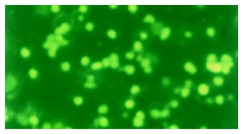
Figure 1 PBL-T with AICD took on kelly fluorescence and the cell without AICD did not take on any fluorescence (TUNEL staining, 200 ×, fluorescence microscope).
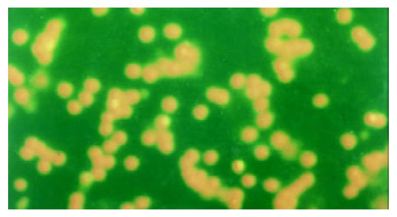
Figure 2 The plasm of PBL-T with AICD took on red fluores-cence and the nucleus took on kelly fluorescence.
But the cell without AICD only took on red fluorescence (TUNEL and PI double staining, fluorescence microscope, 200 ×).
- Citation: Hou CS, Wang GQ, Lu SL, Yue B, Li MR, Wang XY, Yu JW. Role of activation-induced cell death in pathogenesis of patients with chronic hepatitis B. World J Gastroenterol 2003; 9(10): 2356-2358
- URL: https://www.wjgnet.com/1007-9327/full/v9/i10/2356.htm
- DOI: https://dx.doi.org/10.3748/wjg.v9.i10.2356


