Copyright
©The Author(s) 2002.
World J Gastroenterol. Jun 15, 2002; 8(3): 511-514
Published online Jun 15, 2002. doi: 10.3748/wjg.v8.i3.511
Published online Jun 15, 2002. doi: 10.3748/wjg.v8.i3.511
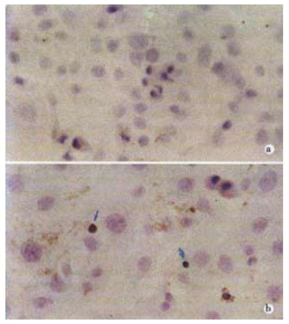
Figure 1 (A) Control HSC (TUNEL × 400).
(B) Apoptotic cell characterized by compaction of nuclear chromatin and condensation of cytoplasm (TUNEL × 400). By TUNEL stain the most condensed cells demonstrated evidence of DNA fragmentation and were strongly staining (iü).
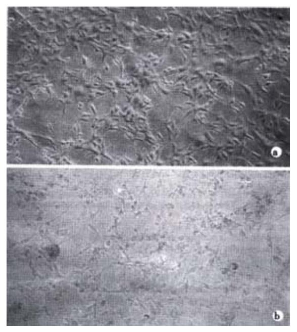
Figure 2 Morphological changes of HSC observed by light microscopy treated by Yigan Decoction.
(A) Untreated cells (× 200); (B) some HSC got round, detached and floating in the culture supernatant after exposure to 18 g·L-1 Yigan Decoction for 48 h (× 100).
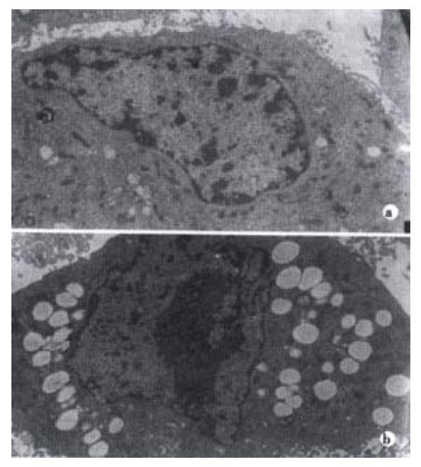
Figure 3 Ultrastructures of HSC with or without treatment with Yigan Decoction.
Following 48 h treatment of Yigan Decoction, dilated endoplasmic reticulum, irregular nuclei, chromatin condensation and heterochromatin ranked along inside of nuclear membrane could be found (A 5000 ×). Untreated cells (B 5000 ×).
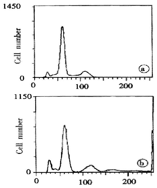
Figure 4 Flow cytometry changes.
(A) Control group. (B) Yigan Decoction group.
- Citation: Yao XX, Tang YW, Yao DM, Xiu HM. Effects of Yigan Decoction on proliferation and apoptosis of hepatic stellate cells. World J Gastroenterol 2002; 8(3): 511-514
- URL: https://www.wjgnet.com/1007-9327/full/v8/i3/511.htm
- DOI: https://dx.doi.org/10.3748/wjg.v8.i3.511


