Copyright
©The Author(s) 2002.
World J Gastroenterol. Feb 15, 2002; 8(1): 153-157
Published online Feb 15, 2002. doi: 10.3748/wjg.v8.i1.153
Published online Feb 15, 2002. doi: 10.3748/wjg.v8.i1.153
Math 1 Math(A1).
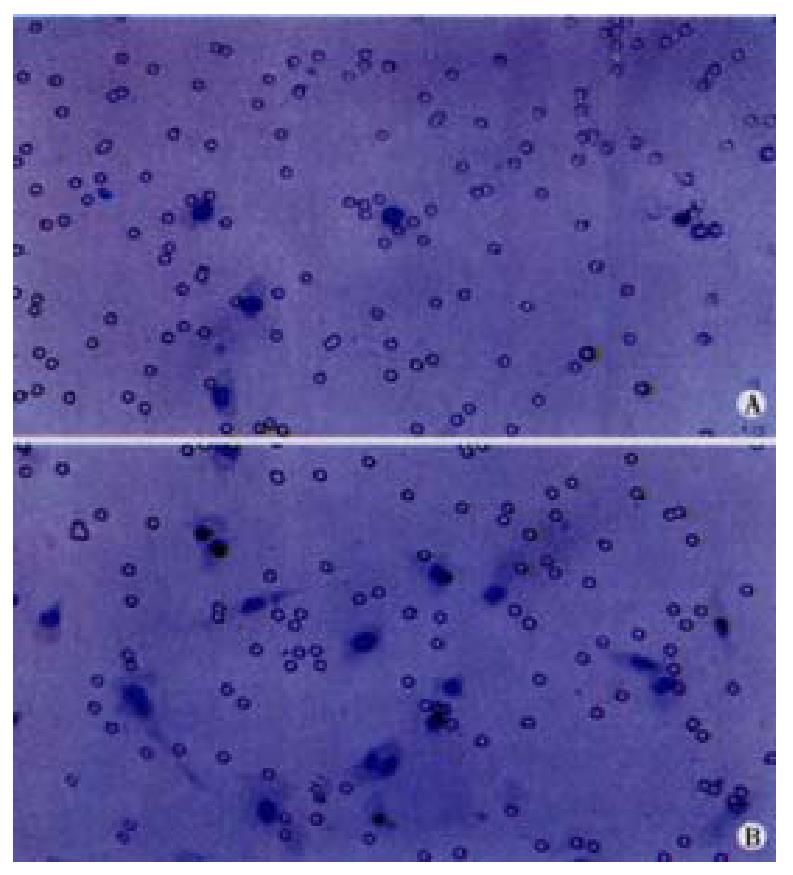
Figure 1 The migration of endothelial cells induced by LS174T cells was increased after the cancer cells were exposed to 10-5 mol·L-1 H2O2.
The regular circles is the 8ìm pores located in PET membrane. A: induced by cancer cells without H2O2 treatment. B: induced by cancer cells with H2O2 treatment. × 200
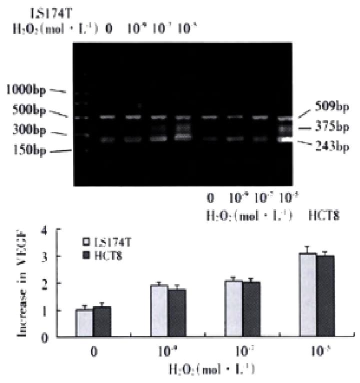
Figure 2 Expression levels of VEGF were increased in LS174T and HCT8 after exposure to 10-9, 10-7 and 10-5 mol·L-1 H2O2, demonstrating a dose-de-pendent feature.
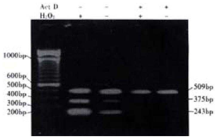
Figure 3 LS174T cells were treated with 1.
5 mg·L-1 Act D for 4 h, followed by exposure to 10-5 mol·L-1 H2O2 for 2 4h. RT-PCR was done. The control was those without Act D treatment.
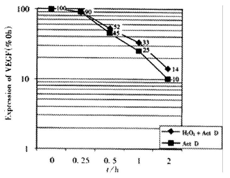
Figure 4 Effect of H2O2 on the half-life of VEGF mRNA in LS174T cells with or without H2O2 exposure following Act D treatment.
No difference in half-life was showed between the two groups.
Figure 5 Measurement of nuclear and cytoplasmic NF-κB associated fluorescence by LSC.
The red fluorescence represents the nuclear area, and the intensity of green fluorescence over nucleus(Fn) reflects NF-κB activity. A, B: LS174T cells without H2O2 treatment. C, D: Increase in NF-κB activity of LS174T cells with H2O2 exposure. E, F: HCT8 cells without H2O2 treatment. G, H: Increase in NF-κB activity of LS174T cells with H2O2 exposure.
- Citation: Zhu JW, Yu BM, Ji YB, Zheng MH, Li DH. Upregulation of vascular endothelial growth factor by hydrogen peroxide in human colon cancer. World J Gastroenterol 2002; 8(1): 153-157
- URL: https://www.wjgnet.com/1007-9327/full/v8/i1/153.htm
- DOI: https://dx.doi.org/10.3748/wjg.v8.i1.153


