Copyright
©The Author(s) 2001.
World J Gastroenterol. Feb 15, 2001; 7(1): 53-59
Published online Feb 15, 2001. doi: 10.3748/wjg.v7.i1.53
Published online Feb 15, 2001. doi: 10.3748/wjg.v7.i1.53
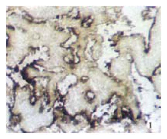
Figure 1 Primary stomach cancer of AFDT with liver metastasis.
AKP was moderately positive and distributed along the free edge of cancerous papillary structure. Frozen section. × 20
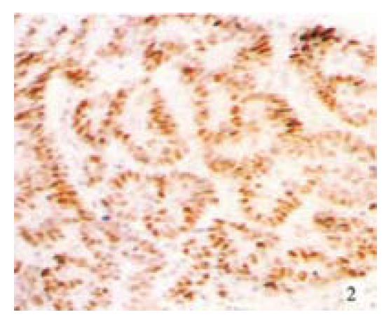
Figure 2 The same case in Figure 1, Tp53 protein was expressed in most of primary cancer cells.
Immunostain, × 16
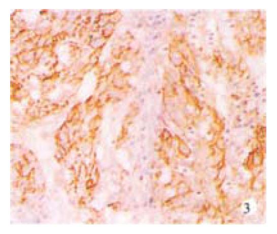
Figure 3 The same case of Figure 1.
CD44v6 was expressed in most of primary cancer cells. Immunostain, × 20
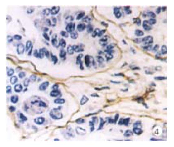
Figure 4 The same case of Figure 1.
There was obvious basement membrane like structure with Laminin positive in the primary tumor. Immunostain, × 40
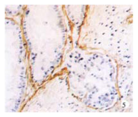
Figure 5 The same case of Figure 1.
There was also obvious basement membrane like structure with laminin positive in the liver metastatic tumor. Immunostain, × 20
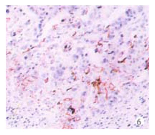
Figure 6 The same case of Figure 1.
CD44v6 was also expressed in the liver metastatic cancer cells. Immunostain, × 20
Figure 7 Primary gastric carcinoma of AMPFDT with ovary metastasis.
LAP was moderately positive and distributed in the membrane and cytoplasm of cancer cells. Frozen section, × 40
Figure 8 The same case of Figure 7.
Sialomucin and neutral mucin were positive in the primary cancer cells. Mucin histochemistry, × 20
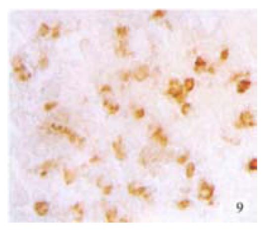
Figure 9 The same case of Figure 7.
Most of primary cancer cells expressed ER, which was distributed in the nuclei and cytoplasm. Immunostain, × 20
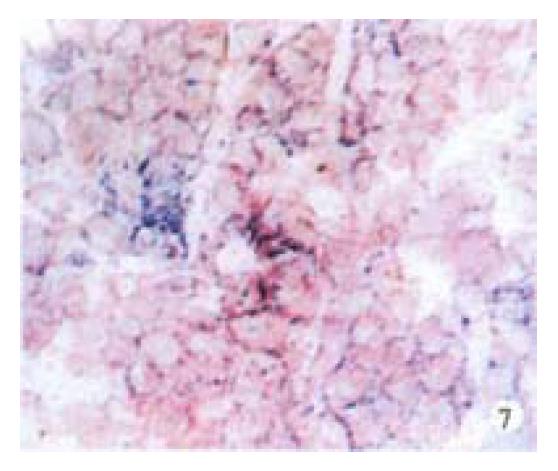
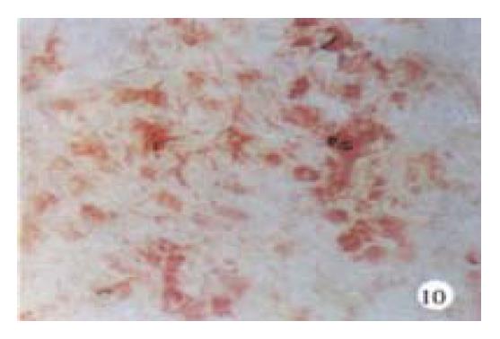
Figure 10 The same case of Figure 7.
LAP was moderately positive in the ovary metastatic cancer cells. Frozen section, × 20
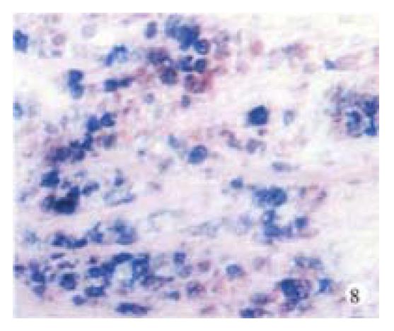
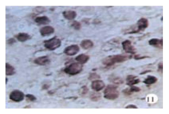
Figure 11 The same case of Figure 7.
Sulfomucin was positive in the ovary metastatic cancer cells. Mucin histochemistry, × 40
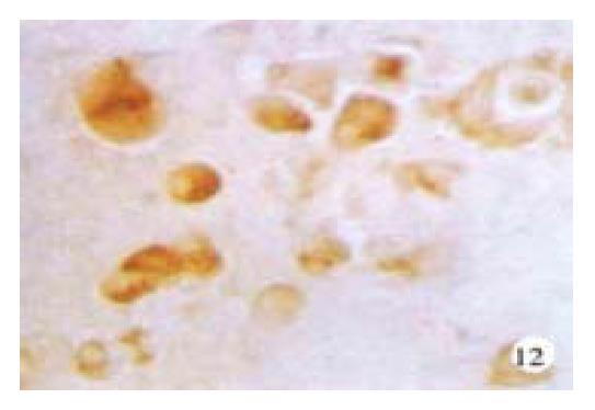
Figure 12 The same case of Figure 7.
Most of the ovary metastatic cancer cells expressed ER. Immunostain, × 40
- Citation: Xin Y, Li XL, Wang YP, Zhang SM, Zheng HC, Wu DY, Zhang YC. Relationship between phenotypes of cell-function differentiation and pathobiological behavior of gastric carcinomas. World J Gastroenterol 2001; 7(1): 53-59
- URL: https://www.wjgnet.com/1007-9327/full/v7/i1/53.htm
- DOI: https://dx.doi.org/10.3748/wjg.v7.i1.53


