Copyright
©The Author(s) 2000.
World J Gastroenterol. Feb 15, 2000; 6(1): 128-130
Published online Feb 15, 2000. doi: 10.3748/wjg.v6.i1.128
Published online Feb 15, 2000. doi: 10.3748/wjg.v6.i1.128
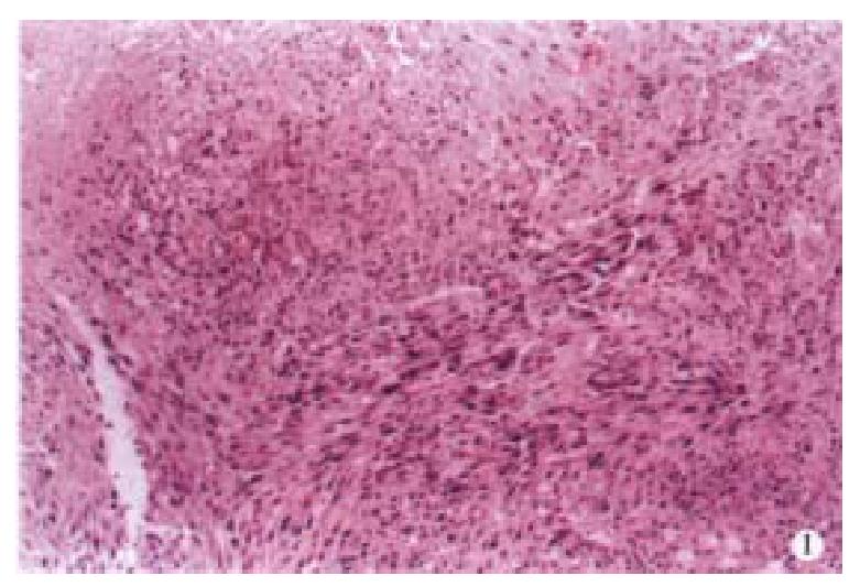
Figure 1 Interlaced arrangement of leiomyosarcoma cells stained with HE.
× 20
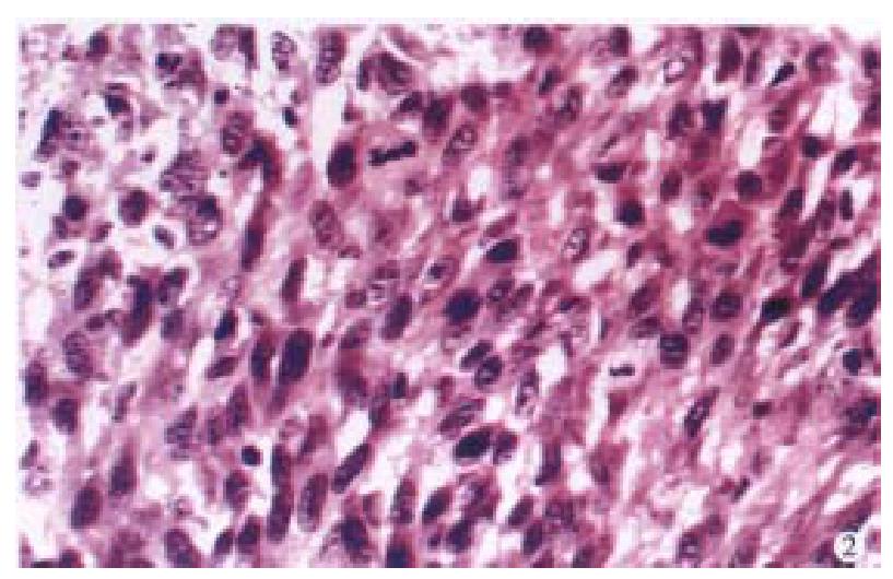
Figure 2 Karyokinesis of leiomyosarcoma cells stained with HE.
× 40
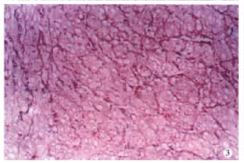
Figure 3 Reticular fibers of leiomyosarcoma cells stained with silver.
× 40
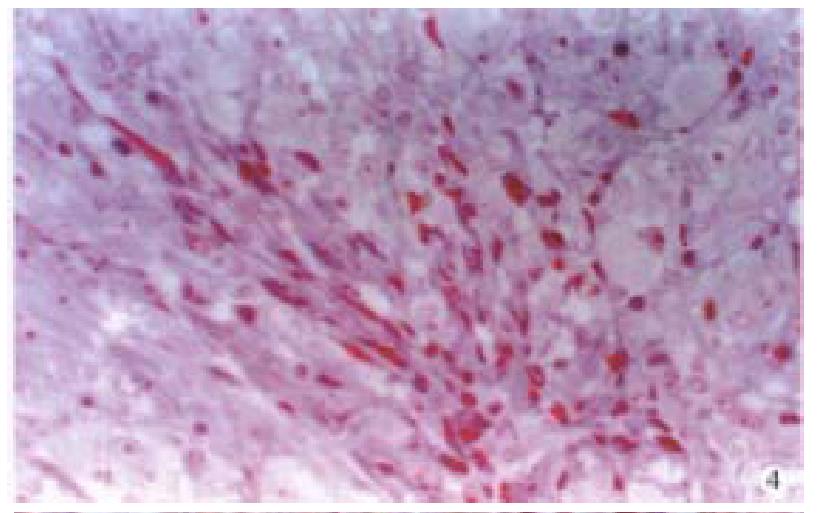
Figure 4 Leiomyosarcoma cells stained red by Masson’s method.
× 40
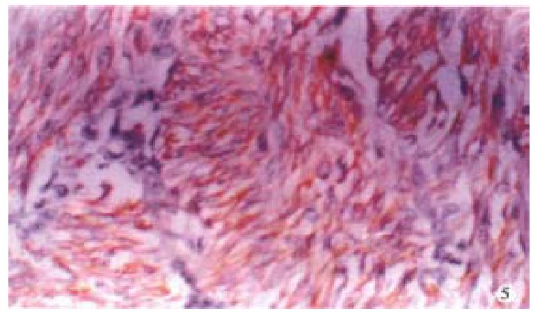
Figure 5 Micrograph of positively stained desmin of cytoplasm of leiomyosarcoma cells by immunohistochemical method.
× 20
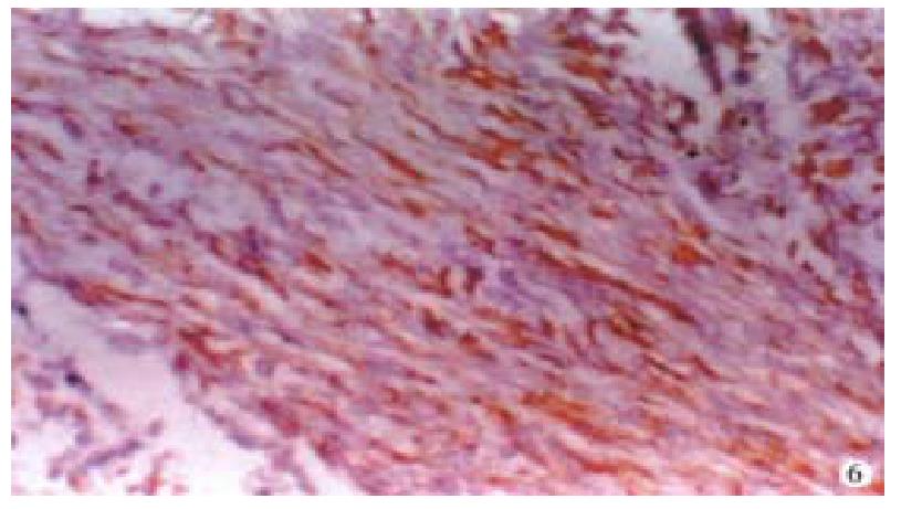
Figure 6 Micrograph of positively stained actin of cytoplasm of leiomyosarcoma cells by immunohistochemical method.
× 20
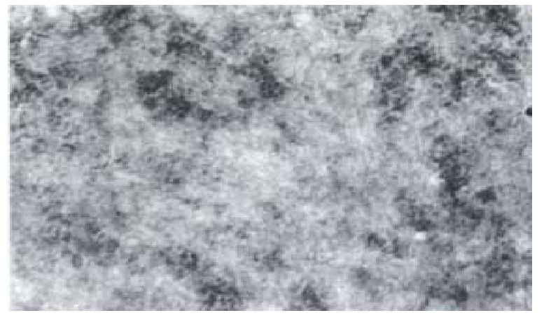
Figure 7 Electron micrograph of dense bodies and myofilaments of leiomyosarcoma.
E.M. × 20000
- Citation: Zhu JS, Su Q, Zhou JG, Hu PL, Xu JH. Study of primary leiomyosarcoma induced by MNNG in BALB/C nude mice. World J Gastroenterol 2000; 6(1): 128-130
- URL: https://www.wjgnet.com/1007-9327/full/v6/i1/128.htm
- DOI: https://dx.doi.org/10.3748/wjg.v6.i1.128


