Copyright
©The Author(s) 1998.
World J Gastroenterol. Oct 15, 1998; 4(5): 392-396
Published online Oct 15, 1998. doi: 10.3748/wjg.v4.i5.392
Published online Oct 15, 1998. doi: 10.3748/wjg.v4.i5.392
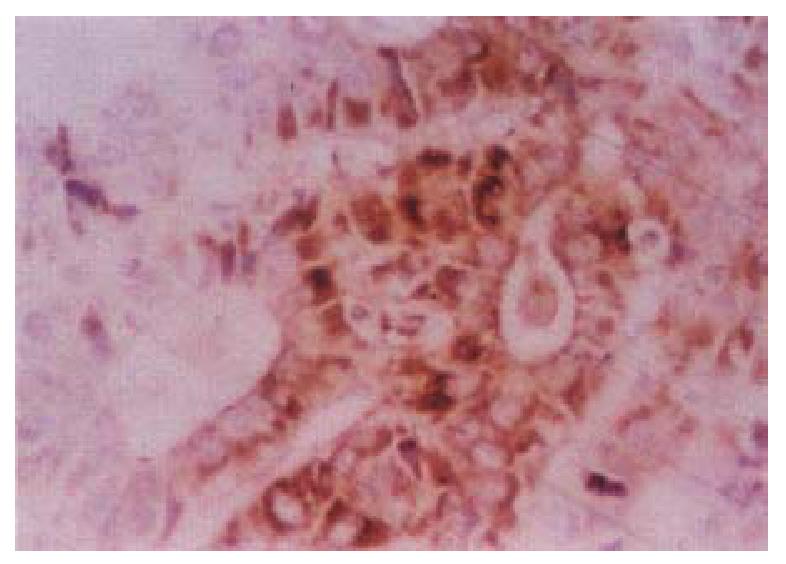
Figure 1 Cancerous surrounding hepatic tissue, HBxAg-positive substance located in the cytoplasms.
ABC × 400
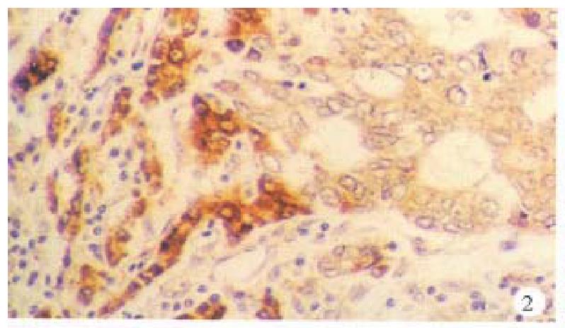
Figure 2 Cancerous and surrounding hepatic tissues, detection rate and intensity of the HBxAg-positive cells in the surrounding hepatic tissues were both higher than that in the cancerous tissues.
ABC × 200
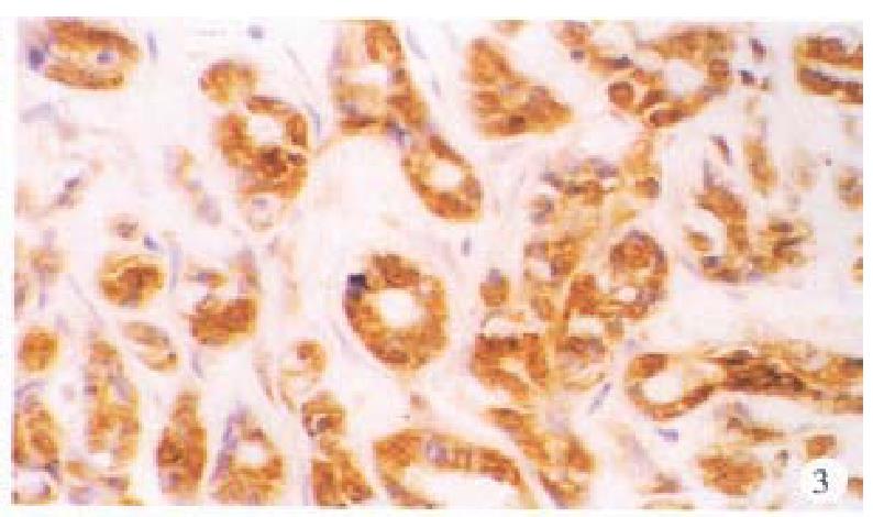
Figure 3 Cancerous tissue, HBsAg-positive substance located mainly in the cytoplasms.
ABC × 200
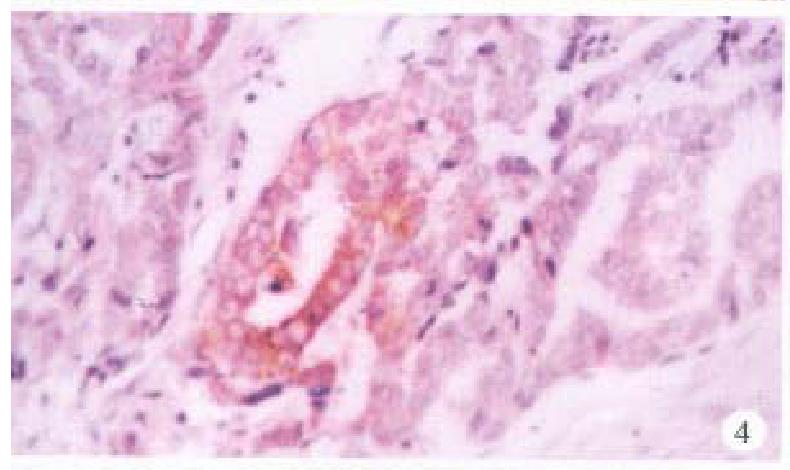
Figure 4 Cancerous tissus, pre-S1 antigens located mainly in the cytoplasms, some on cellular membranes.
ABC × 400
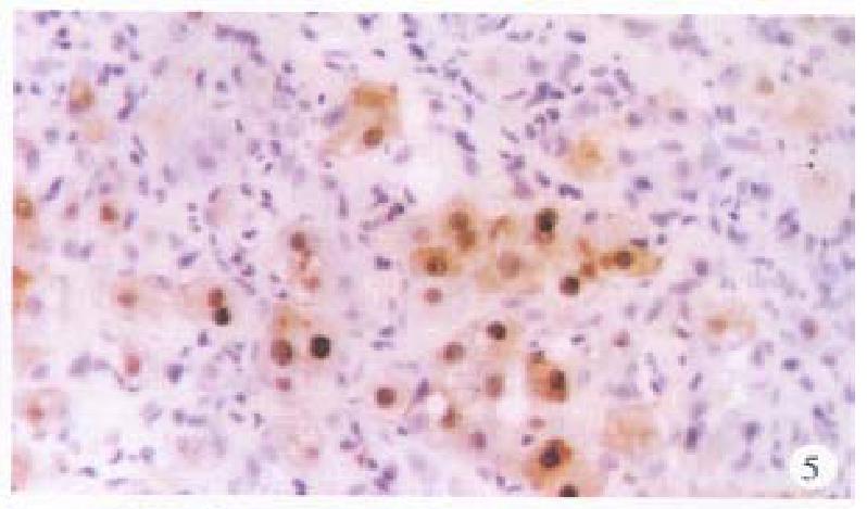
Figure 5 Surrounding hepatic tissue, HBc-Ag positive located mainly in the nuclei, some in the cytoplasms and on cellular membranes.
ABC × 400
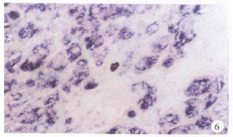
Figure 6 Surrounding hepatic tissue, HBV-DNA gene located in the cytoplasms, some in the nuclei.
ISH × 400
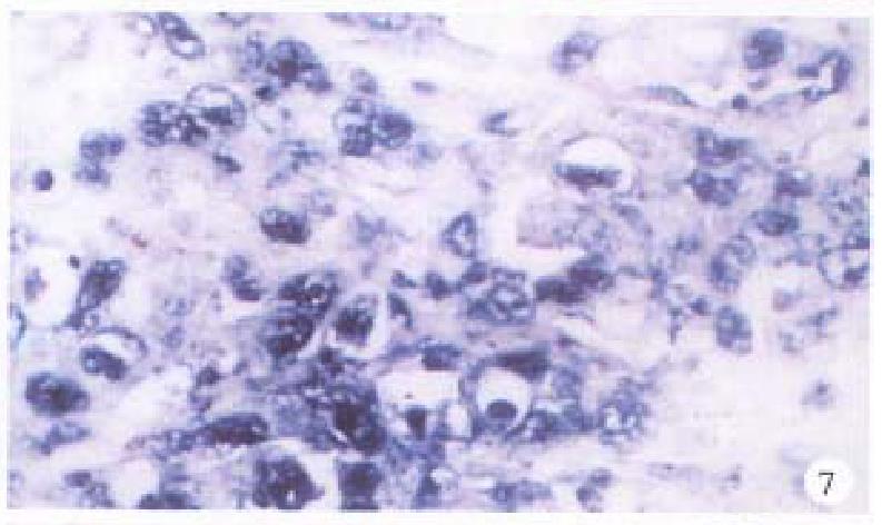
Figure 7 Surrounding hepatic tissue, pre-S gene located in the cytoplasms, some in the nuclei.
ISH × 400
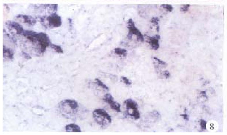
Figure 8 Surrounding hepatic tissue, S gene located mainly in the cytoplasms.
ISH × 400
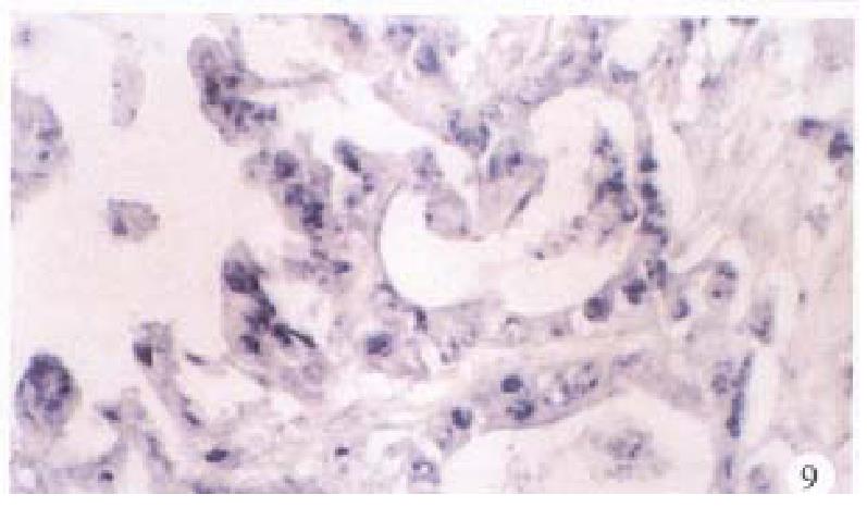
Figure 9 Cancerous tissue, C gene located mainly in the nuclei, some in the cytoplasms.
ISH × 400
- Citation: Wang WL, Gu GY, Hu M. Expression and significance of HBV genes and their antigens in human primary intrahepatic cholangiocarcinoma. World J Gastroenterol 1998; 4(5): 392-396
- URL: https://www.wjgnet.com/1007-9327/full/v4/i5/392.htm
- DOI: https://dx.doi.org/10.3748/wjg.v4.i5.392


