Copyright
©The Author(s) 1997.
World J Gastroenterol. Sep 15, 1997; 3(3): 174-176
Published online Sep 15, 1997. doi: 10.3748/wjg.v3.i3.174
Published online Sep 15, 1997. doi: 10.3748/wjg.v3.i3.174
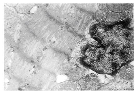
Figure 1 Electron micrograph of rat myocardium from the sham operated (SO) group (× 14000).
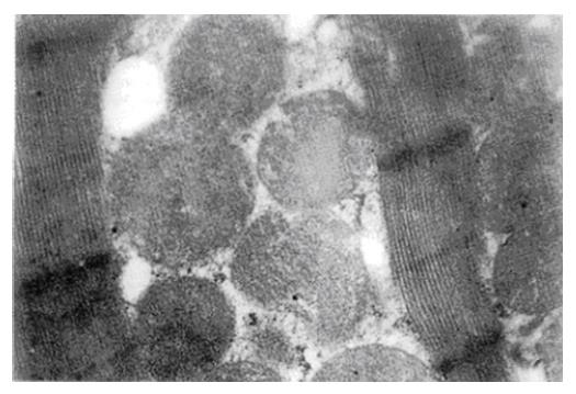
Figure 2 Electron micrograph of rat myocardium from the BDL1 group.
The mitochondria appear swollen, with indistinct outer membranes and obscured cristae (× 21000).
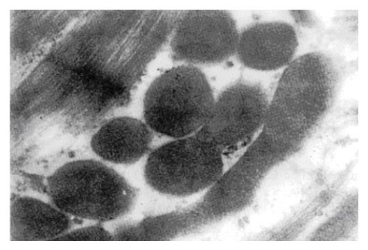
Figure 3 Electron micrograph of rat myocardium from the BDL2 group.
Mitochondria appear distorted and curdled, with indistinct outer membranes and cristae (× 21000).
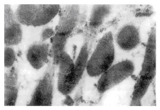
Figure 4 Electron micrograph of myocardium from sodium cholate-fed rats.
The number of mitochondria are decreased and appear distorted, with indistinct outer membranes and cristae (× 21000).
- Citation: Mu YP, Peng SY. Relation between bile acids and myocardial damage in obstructive jaundice. World J Gastroenterol 1997; 3(3): 174-176
- URL: https://www.wjgnet.com/1007-9327/full/v3/i3/174.htm
- DOI: https://dx.doi.org/10.3748/wjg.v3.i3.174


